Dental implant, Radiation therapy, Osteoradionecrosis
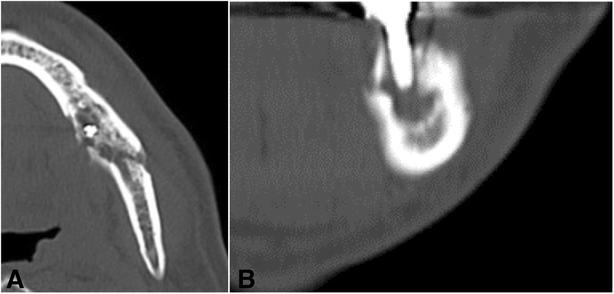
Fig. 3. CT images of the left mandible. a Axial view at the left first molar. b Coronal view at the left first molar : A case of peri-implant
author: Yuji Teramoto, Hiroshi Kurita, Takahiro Kamata, Hitoshi Aizawa, Nobuhiko Yoshimura, Humihiro Nishimaki, Kazunobu Takamizawa | publisher: drg. Andreas Tjandra, Sp. Perio, FISID

Fig. 3. CT images of the left mandible. a Axial view at the left first molar. b Coronal view at the left first molar
Serial posts:
- Abstract : A case of peri-implantitis and osteoradionecrosis arising around dental implants placed before radiation therapy
- Background : A case of peri-implantitis and osteoradionecrosis arising around dental implants placed before radiation therapy
- Case presentation : A case of peri-implantitis and osteoradionecrosis arising around dental implants placed before radiation therapy [1]
- Case presentation : A case of peri-implantitis and osteoradionecrosis arising around dental implants placed before radiation therapy [2]
- Case presentation : A case of peri-implantitis and osteoradionecrosis arising around dental implants placed before radiation therapy [3]
- Conclusions : A case of peri-implantitis and osteoradionecrosis arising around dental implants placed before radiation therapy
- Consent : A case of peri-implantitis and osteoradionecrosis arising around dental implants placed before radiation therapy
- References : A case of peri-implantitis and osteoradionecrosis arising around dental implants placed before radiation therapy
- Author information : A case of peri-implantitis and osteoradionecrosis arising around dental implants placed before radiation therapy
- Additional information : A case of peri-implantitis and osteoradionecrosis arising around dental implants placed before radiation therapy
- Rights and permissions : A case of peri-implantitis and osteoradionecrosis arising around dental implants placed before radiation therapy
- About this article : A case of peri-implantitis and osteoradionecrosis arising around dental implants placed before radiation therapy
- Fig. 1. Intraoral photo at the first visit : A case of peri-implant
- Fig. 2. Panoramic X-ray image at the first visit : A case of peri-implant
- Fig. 3. CT images of the left mandible. a Axial view at the left first molar. b Coronal view at the left first molar : A case of peri-implant
- Fig. 4. a Intraoperative photo. The affected left mandible was segmentally resected. b Intraoperative photo. A vascularized fibula bone graft. c Resected mandible. d Panoramic X-ray image after the surgery : A case of peri-implant
- Fig. 5. Histopathologic photo of the resected mandible (H-E staining) : A case of peri-implant
- Fig. 6. a Panoramic X-ray image 1 year after the surgery. b Intraoral photo 1 year after the surgery : A case of peri-implant