Ridge Preservation, Bone Graft Substitutes (BGS), Cone Beam CT (CBCT), Socket Grafting, Ridge Width
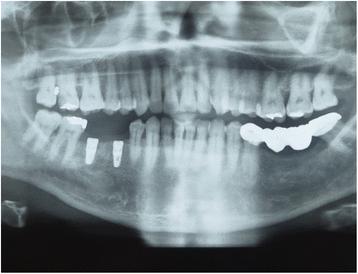
Fig. 3. Postoperative X ray showing the implant positions in the mandible where the teeth were extracted and ridge preservation was accomplished : Ridge preservation using an in situ hardening biph
author: Ashish Kakar, Bappanadu H Sripathi Rao, Shashikanth Hegde, Nikhil Deshpande, Annette Lindner, Heiner Nagursky, Aditya Patney, Ha | publisher: drg. Andreas Tjandra, Sp. Perio, FISID

Fig. 3. Postoperative X ray showing the implant positions in the mandible where the teeth were extracted and ridge preservation was accomplished
Serial posts:
- Abstract : Ridge preservation using an in situ hardening biphasic calcium phosphate (β-TCP/HA) bone graft substitute—a clinical, radiological, and histological study
- Background : Ridge preservation using an in situ hardening biphasic calcium phosphate (β-TCP/HA) bone graft substitute—a clinical, radiological, and histological study [1]
- Background : Ridge preservation using an in situ hardening biphasic calcium phosphate (β-TCP/HA) bone graft substitute—a clinical, radiological, and histological study [2]
- Methods : Ridge preservation using an in situ hardening biphasic calcium phosphate (β-TCP/HA) bone graft substitute—a clinical, radiological, and histological study [1]
- Methods : Ridge preservation using an in situ hardening biphasic calcium phosphate (β-TCP/HA) bone graft substitute—a clinical, radiological, and histological study [2]
- Methods : Ridge preservation using an in situ hardening biphasic calcium phosphate (β-TCP/HA) bone graft substitute—a clinical, radiological, and histological study [3]
- Results : Ridge preservation using an in situ hardening biphasic calcium phosphate (β-TCP/HA) bone graft substitute—a clinical, radiological, and histological study [1]
- Results : Ridge preservation using an in situ hardening biphasic calcium phosphate (β-TCP/HA) bone graft substitute—a clinical, radiological, and histological study [2]
- Discussion : Ridge preservation using an in situ hardening biphasic calcium phosphate (β-TCP/HA) bone graft substitute—a clinical, radiological, and histological study [1]
- Discussion : Ridge preservation using an in situ hardening biphasic calcium phosphate (β-TCP/HA) bone graft substitute—a clinical, radiological, and histological study [2]
- Discussion : Ridge preservation using an in situ hardening biphasic calcium phosphate (β-TCP/HA) bone graft substitute—a clinical, radiological, and histological study [3]
- Conclusions : Ridge preservation using an in situ hardening biphasic calcium phosphate (β-TCP/HA) bone graft substitute—a clinical, radiological, and histological study
- References : Ridge preservation using an in situ hardening biphasic calcium phosphate (β-TCP/HA) bone graft substitute—a clinical, radiological, and histological study [1]
- References : Ridge preservation using an in situ hardening biphasic calcium phosphate (β-TCP/HA) bone graft substitute—a clinical, radiological, and histological study [2]
- References : Ridge preservation using an in situ hardening biphasic calcium phosphate (β-TCP/HA) bone graft substitute—a clinical, radiological, and histological study [3]
- References : Ridge preservation using an in situ hardening biphasic calcium phosphate (β-TCP/HA) bone graft substitute—a clinical, radiological, and histological study [4]
- Acknowledgements : Ridge preservation using an in situ hardening biphasic calcium phosphate (β-TCP/HA) bone graft substitute—a clinical, radiological, and histological study
- Author information : Ridge preservation using an in situ hardening biphasic calcium phosphate (β-TCP/HA) bone graft substitute—a clinical, radiological, and histological study [1]
- Author information : Ridge preservation using an in situ hardening biphasic calcium phosphate (β-TCP/HA) bone graft substitute—a clinical, radiological, and histological study [2]
- Rights and permissions : Ridge preservation using an in situ hardening biphasic calcium phosphate (β-TCP/HA) bone graft substitute—a clinical, radiological, and histological study
- About this article : Ridge preservation using an in situ hardening biphasic calcium phosphate (β-TCP/HA) bone graft substitute—a clinical, radiological, and histological study
- Table 1 Buccal and palatal ISQ values at implant placement and time of loading : Ridge preservation using an in situ hardening biphasic calcium phosphate (β-TCP/HA) bone graft substitute—a clinical, radiological, and histological study
- Table 2 Width ridge changes assess by cone beam computer tomography (CBCT) : Ridge preservation using an in situ hardening biphasic calcium phosphate (β-TCP/HA) bone graft substitute—a clinical, radiological, and histological study
- Table 3 Histomorphometric evaluation of core biopsy sections : Ridge preservation using an in situ hardening biphasic calcium phosphate (β-TCP/HA) bone graft substitute—a clinical, radiological, and histological study
- Fig. 1. a Clinical occlusal view with fractured 45 and 46. b Post-extraction view of the socket. Note minimal trauma to the soft tissue and no flap reflection on the surgical site. c Graft material condensed into the extraction sockets showing good initial graft stability. d Black silk sutures placed with tissue approximation and no releasing incision in the flaps : Ridge preservation using an in situ hardening biph
- Fig. 2. a Clinical postoperative view after 4 months. Note that the healing was achieved only with tissue approximation. A good width of keratinized tissue is visible along with ridge preservation. Ready for implant placement in the grafted areas. b Implant placed in 45 area. Core biopsy sample taken from area 46. Note the integration of graft particles in the preserved alveolar ridge also inside the osteotomy site of 46. c Two Xive (Dentsply) implants placed in the preserved ridge. d. Postoperative X ray showing the implant positions in the mandible where the teeth were extracted and ridge preservation was accomplished : Ridge preservation using an in situ hardening biph
- Fig. 3. Postoperative X ray showing the implant positions in the mandible where the teeth were extracted and ridge preservation was accomplished : Ridge preservation using an in situ hardening biph
- Fig. 4. a Second stage surgery followed by impression making. Note the excellent width of keratinized tissue which was also preserved. b Implant crowns placed and loaded after 3 months of placement : Ridge preservation using an in situ hardening biph
- Fig. 5. CBCT images of the extraction site. a Preoperative CBCT showing fractured and un-restorable teeth #45 and #46 planned to be extracted. b–d Cross sectional views : Ridge preservation using an in situ hardening biph
- Fig. 6. a–c Four-month postoperative CBCT showing graft integration and preservation of ridge without collapse of the buccal or lingual cortical plates also showing the cross sections in the grafted area : Ridge preservation using an in situ hardening biph
- Fig. 7. a–c Histological sections of bone core biopsy taken from the site of implantation using a trephine bur. a Overview image of coronal-apical cut through the entire core biopsy showing formation of new bone (NB) next to old bone of the extraction socket (B). easy-graft CRYSTAL particles (Gr) are embedded in well perfused connective tissue (CT) and new bone (NB) (Azur II and Pararosanilin, original magnification ×50). b Integration of easy-graft CRYSTAL particle (Gr) into newly formed bone (NB) and connective tissue (CT) showing tight contact between graft particle and new bone. c High magnification (×200) images of the interface between graft particle (Gr) and new bone (NB) showing osteoblasts (OB) forming osteoid (O) and formation of new bone (NB) on the surface of easy-graft CRYSTAL particles (Gr) (Azur II and Pararosanilin, original magnification ×200) : Ridge preservation using an in situ hardening biph