Dental implant, Screw-retained, Access-hole, Wear, 4-META
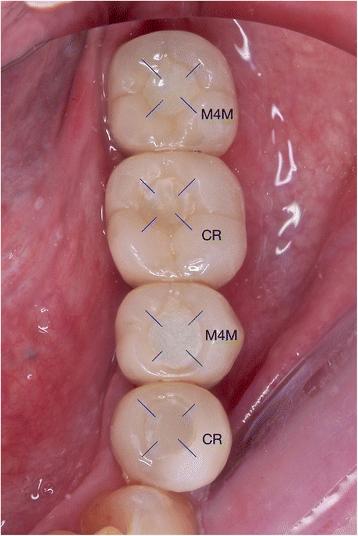
Fig. 4. Margin depth measurement localization (example: TRA, T = 12 M) : Comparison of access-hole filling materials for screw retained implant
author: Rmy Tanimura, Shiro Suzuki | publisher: drg. Andreas Tjandra, Sp. Perio, FISID

Fig. 4. Margin depth measurement localization (example: TRA, T = 12 M)
Serial posts:
- Abstract : Comparison of access-hole filling materials for screw retained implant prostheses: 12-month in vivo study
- Background : Comparison of access-hole filling materials for screw retained implant prostheses: 12-month in vivo study
- Methods : Comparison of access-hole filling materials for screw retained implant prostheses: 12-month in vivo study [1]
- Methods : Comparison of access-hole filling materials for screw retained implant prostheses: 12-month in vivo study [2]
- Methods : Comparison of access-hole filling materials for screw retained implant prostheses: 12-month in vivo study [3]
- Results : Comparison of access-hole filling materials for screw retained implant prostheses: 12-month in vivo study
- Discussion : Comparison of access-hole filling materials for screw retained implant prostheses: 12-month in vivo study [3]
- References : Comparison of access-hole filling materials for screw retained implant prostheses: 12-month in vivo study [1]
- References : Comparison of access-hole filling materials for screw retained implant prostheses: 12-month in vivo study [2]
- Table 1 ᅟ : Comparison of access-hole filling materials for screw retained implant prostheses: 12-month in vivo study
- Table 2 Aesthetical Outcomes at T = 12 M (VAS Score) : Comparison of access-hole filling materials for screw retained implant prostheses: 12-month in vivo study
- Table 3 Surface areas changes of access-hole filling. Unit: % : Comparison of access-hole filling materials for screw retained implant prostheses: 12-month in vivo study
- Table 4 Disappearance of the overfilling. Unit: % : Comparison of access-hole filling materials for screw retained implant prostheses: 12-month in vivo study
- Fig. 1. Brush-dip technique : Comparison of access-hole filling materials for screw retained implant
- Fig. 2. Occlusal contact point : Comparison of access-hole filling materials for screw retained implant
- Fig. 3. a–e (Filling surface changes): a (ROG, T = 0). b (ROG, T = 1 M). c (ROG, T = 3 M). d (ROG, T = 6 M). e (ROG, T = 12 M) : Comparison of access-hole filling materials for screw retained implant
- Fig. 4. Margin depth measurement localization (example: TRA, T = 12 M) : Comparison of access-hole filling materials for screw retained implant
- Fig. 5. Depth and angle at the margin : Comparison of access-hole filling materials for screw retained implant
- Fig. 6. Access-hole filling surface areas measurement, average : Comparison of access-hole filling materials for screw retained implant
- Fig. 7. a, b (The marginal discrepancy pattern for group CR and M4M). a Group CR (1: Ceramic surface, 2: CR surface) Units of the axis are in μm. b Group M4M (1: Ceramic surface, 2: M4M surface) Units of the axis are in μm : Comparison of access-hole filling materials for screw retained implant