Alveolar ridge preservation, Particulated dentin, Autologous augmentation, Bone augmentation, Bone substitute
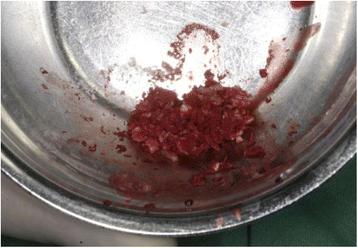
Fig. 7. Autologous, particulated dentin mixed with blood from the operating site : Alveolar ridge preservation with autologous partic
author: Silvio Valdec, Pavla Pasic, Alex Soltermann, Daniel Thoma, Bernd Stadlinger, Martin Rcker | publisher: drg. Andreas Tjandra, Sp. Perio, FISID

Fig. 7. Autologous, particulated dentin mixed with blood from the operating site
Serial posts:
- Background : Alveolar ridge preservation with autologous particulated dentin—a case series
- Material and methods : Alveolar ridge preservation with autologous particulated dentin—a case series [1]
- Material and methods : Alveolar ridge preservation with autologous particulated dentin—a case series [2]
- Case presentation : Alveolar ridge preservation with autologous particulated dentin—a case series
- Results : Alveolar ridge preservation with autologous particulated dentin—a case series
- Discussion : Alveolar ridge preservation with autologous particulated dentin—a case series [1]
- Discussion : Alveolar ridge preservation with autologous particulated dentin—a case series [2]
- Conclusion : Alveolar ridge preservation with autologous particulated dentin—a case series
- References : Alveolar ridge preservation with autologous particulated dentin—a case series [1]
- References : Alveolar ridge preservation with autologous particulated dentin—a case series [2]
- References : Alveolar ridge preservation with autologous particulated dentin—a case series [3]
- References : Alveolar ridge preservation with autologous particulated dentin—a case series [4]
- Acknowledgements : Alveolar ridge preservation with autologous particulated dentin—a case series
- Author information : Alveolar ridge preservation with autologous particulated dentin—a case series
- Rights and permissions : Alveolar ridge preservation with autologous particulated dentin—a case series
- About this article : Alveolar ridge preservation with autologous particulated dentin—a case series
- Fig. 1. Extraction with the benex system : Alveolar ridge preservation with autologous partic
- Fig. 2. The remaining root of tooth 11 : Alveolar ridge preservation with autologous partic
- Fig. 3. Removal of the pulp : Alveolar ridge preservation with autologous partic
- Fig. 4. Removal of enamel and the cementum : Alveolar ridge preservation with autologous partic
- Fig. 5. Autologous dentin in a bone mill : Alveolar ridge preservation with autologous partic
- Fig. 6. Autologous dentin with the desired particle size : Alveolar ridge preservation with autologous partic
- Fig. 7. Autologous, particulated dentin mixed with blood from the operating site : Alveolar ridge preservation with autologous partic
- Fig. 8. Autologous, particulated dentin in the alveolar socket : Alveolar ridge preservation with autologous partic
- Fig. 9. Soft tissue punch : Alveolar ridge preservation with autologous partic
- Fig. 10. Soft tissue graft placed on the recipient site : Alveolar ridge preservation with autologous partic
- Fig. 11. Sagittal view : Alveolar ridge preservation with autologous partic
- Fig. 12. Axial view : Alveolar ridge preservation with autologous partic
- Fig. 13. a, b Clinical situation prior to implant placement : Alveolar ridge preservation with autologous partic
- Fig. 14. Single tooth X-ray immediately after the augmentation using autogenous dentin : Alveolar ridge preservation with autologous partic
- Fig. 15. Single tooth X-ray, showing a constant bone level 7 months after implant placement : Alveolar ridge preservation with autologous partic
- Fig. 16. Single tooth X-ray, 1 year post-implantation, showing the finalized crown : Alveolar ridge preservation with autologous partic
- Fig. 17. Histology of dentin augmentation. aAsterisk denotes incorporated dentin particle, surrounded by vital woven bone. Triangle shows reactive process in the bone marrow lacunae with osteoblast rimming. No signs of necrosis or infection (H&E stain, ×100 magnification). b Larger magnification at ×200. c EvG (Elastica van Gieson) stain, ×200 : Alveolar ridge preservation with autologous partic
- Fig. 18. Finalized prosthetic restoration after 1 year : Alveolar ridge preservation with autologous partic
- Fig. 19. Colour-coded superimposition of intraoral scans before extraction and after definitive prosthetic restoration : Alveolar ridge preservation with autologous partic
- Fig. 20. Colour-coded superimposition of intraoral scans before extraction and after definitive prosthetic restoration : Alveolar ridge preservation with autologous partic