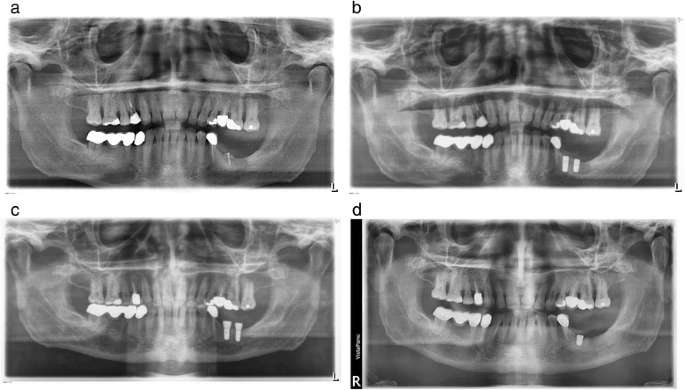
Figure 1. a Patient 1. Post grafting orthopantomogram. The bone block was secured with a single microscrew. b Patient 1. The radiograph demonstrates veritable inserted Straumann bone level implants after the first implant placement (1 day after implant placement). A peri-implant osteolysis is not visible. c Patient 1. Postoperative orthopantomogram (1 day after implant placement) after second implant placement of Straumann tissue level implants 6 months later. A peri-implant osteolysis is not visible. d Patient 1. Postoperative orthopantomogram after third implant placement (Conelog ScrewLine implant)
Figure 1. a Patient 1. Post grafting orthopantomogram
author: Tobias Fretwurst,Sebastian Grunert,Johan P Woelber,Katja Nelson,Wiebke Semper-Hogg | publisher: drg. Andreas Tjandra, Sp. Perio, FISID

Serial posts:
- Vitamin D deficiency in early implant failure: two case reports
- Background : Vitamin D deficiency in early implant failure: two case reports
- Case presentation : Vitamin D deficiency in early implant failure (1)
- Case presentation : Vitamin D deficiency in early implant failure (2)
- Case presentation : Vitamin D deficiency in early implant failure (3)
- Discussion : Vitamin D deficiency in early implant failure (1)
- Discussion : Vitamin D deficiency in early implant failure (2)
- Discussion : Vitamin D deficiency in early implant failure (3)
- References : Vitamin D deficiency in early implant failure
- Figure 1. a Patient 1. Post grafting orthopantomogram
- Figure 2. a Patient 2. Postoperative orthopantomogram one day after implant
- Table 1 Implant characteristics—insertion region/explantation