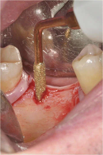
Figure 13. Implant site preparation: OP5, IM2, OT4, and IM3 (correctly in sequence)
Figure 13. Implant site preparation: OP5, IM2, OT4, and IM3 (correctly in sequence)
author: Federico Gelpi,Daniele De Santis,Simone Marconcini,Francesco Briguglio, Marco Finotti | publisher: drg. Andreas Tjandra, Sp. Perio, FISID

Serial posts:
- A piezo surgery with corticotomies and implant placement
- A piezo surgery with corticotomies and implant placement (1)
- A piezo surgery with corticotomies and implant placement (2)
- Discussion : A piezo surgery with corticotomies and implant placement (1)
- Discussion : A piezo surgery with corticotomies and implant placement (2)
- Figure 1. Initial frontal intraoral aspect
- Figure 2. Initial lateral intraoral aspect
- Figure 3. Some metal ceramic crowns in the upper left maxillary arch with a very poor esthetic appearance
- Figure 4. The panoramic radiography and cephalometric analysis revealed a partially edentulous mandible
- Figure 5. The panoramic radiography and cephalometric analysis revealed a partially edentulous mandible
- Figure 6. Orthodontic bracket placement: frontal view
- Figure 7. Ortodontic bracket placement: right side view
- Figure 8. Orthodontic bracket placement: left side view
- Figure 9. A microsurgical corticotomy was mandatory to assist orthodontic tipping and intrusion of elements 16 and 17
- Figure 10. A triangular-shaped corticotomy was performed
- Figure 11. A mesiobuccal root surface exposure of element
- Figure 12. The total width flap was sutured
- Figure 13. Implant site preparation: OP5, IM2, OT4, and IM3 (correctly in sequence)
- Figure 14. Implants placement after site preparation
- Figure 15. All implants received immediate healing screws
- Figure 16. After orthodontic treatment was completed, the prosthodontic phase took place
- Figure 17. Implants were used for implant-retained prostheses (abutment-cemented crowns), and a three-unit fixed partial denture pontic (crowns 25–27) was placed
- Figure 18. OPT after prosthodontic finalization
- Figure 19. A full-mouth frontal aspect
- Figure 20. The left side could not be restored to an ideal class I relationship
- Figure 21. A dental class I occlusion was established only on the right side