Sinus floor elevation, Xenograft, Autogenous bone, Collagen membrane, Osteoconductivity
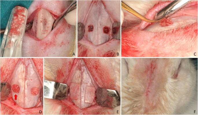
Fig. 1. Clinical view of the surgical procedures. a Tibial bone exposed for autogenous bone harvesting using a bone scraper. b Antrostomies prepared. c Autogenous bone particles placed in the antrostomy. d Xenograft and bone particles (red arrow) at the antrostomies. e Collagen membranes placed on the antrostomies. f Wounds closed with sutures : Influence of the use of autogenous bone particles
author: Giacomo Favero, Jose Via-Almunia, Carmen Carda, Jos Javier Martn de Llano, Berta Garca-Mira, David Soto-Pealoza, Miguel Pearroch | publisher: drg. Andreas Tjandra, Sp. Perio, FISID

Fig. 1. Clinical view of the surgical procedures. a Tibial bone exposed for autogenous bone harvesting using a bone scraper. b Antrostomies prepared. c Autogenous bone particles placed in the antrostomy. d Xenograft and bone particles (red arrow) at the antrostomies. e Collagen membranes placed on the antrostomies. f Wounds closed with sutures
Serial posts:
- Abstract : Influence of the use of autogenous bone particles to close the access window after maxillary sinus floor augmentation: an experimental study in rabbits
- Introduction : Influence of the use of autogenous bone particles to close the access window after maxillary sinus floor augmentation: an experimental study in rabbits [1]
- Introduction : Influence of the use of autogenous bone particles to close the access window after maxillary sinus floor augmentation: an experimental study in rabbits [2]
- Materials and methods : Influence of the use of autogenous bone particles to close the access window after maxillary sinus floor augmentation: an experimental study in rabbits [1]
- Materials and methods : Influence of the use of autogenous bone particles to close the access window after maxillary sinus floor augmentation: an experimental study in rabbits [2]
- Materials and methods : Influence of the use of autogenous bone particles to close the access window after maxillary sinus floor augmentation: an experimental study in rabbits [3]
- Results : Influence of the use of autogenous bone particles to close the access window after maxillary sinus floor augmentation: an experimental study in rabbits [1]
- Results : Influence of the use of autogenous bone particles to close the access window after maxillary sinus floor augmentation: an experimental study in rabbits [2]
- Discussion : Influence of the use of autogenous bone particles to close the access window after maxillary sinus floor augmentation: an experimental study in rabbits [1]
- Discussion : Influence of the use of autogenous bone particles to close the access window after maxillary sinus floor augmentation: an experimental study in rabbits [2]
- Discussion : Influence of the use of autogenous bone particles to close the access window after maxillary sinus floor augmentation: an experimental study in rabbits [3]
- Availability of data and materials : Influence of the use of autogenous bone particles to close the access window after maxillary sinus floor augmentation: an experimental study in rabbits
- Abbreviations : Influence of the use of autogenous bone particles to close the access window after maxillary sinus floor augmentation: an experimental study in rabbits
- References : Influence of the use of autogenous bone particles to close the access window after maxillary sinus floor augmentation: an experimental study in rabbits [1]
- References : Influence of the use of autogenous bone particles to close the access window after maxillary sinus floor augmentation: an experimental study in rabbits [2]
- References : Influence of the use of autogenous bone particles to close the access window after maxillary sinus floor augmentation: an experimental study in rabbits [3]
- References : Influence of the use of autogenous bone particles to close the access window after maxillary sinus floor augmentation: an experimental study in rabbits [4]
- Funding : Influence of the use of autogenous bone particles to close the access window after maxillary sinus floor augmentation: an experimental study in rabbits
- Author information : Influence of the use of autogenous bone particles to close the access window after maxillary sinus floor augmentation: an experimental study in rabbits [1]
- Author information : Influence of the use of autogenous bone particles to close the access window after maxillary sinus floor augmentation: an experimental study in rabbits [2]
- Ethics declarations : Influence of the use of autogenous bone particles to close the access window after maxillary sinus floor augmentation: an experimental study in rabbits
- Additional information : Influence of the use of autogenous bone particles to close the access window after maxillary sinus floor augmentation: an experimental study in rabbits
- Rights and permissions : Influence of the use of autogenous bone particles to close the access window after maxillary sinus floor augmentation: an experimental study in rabbits
- About this article : Influence of the use of autogenous bone particles to close the access window after maxillary sinus floor augmentation: an experimental study in rabbits
- Table 1 Histomorphometric analysis. Tissues evaluated in the various regions after 1 week of healing : Influence of the use of autogenous bone particles to close the access window after maxillary sinus floor augmentation: an experimental study in rabbits
- Table 2 Histomorphometric analysis. Tissues evaluated in the various regions after 8 weeks of healing : Influence of the use of autogenous bone particles to close the access window after maxillary sinus floor augmentation: an experimental study in rabbits
- Fig. 1. Clinical view of the surgical procedures. a Tibial bone exposed for autogenous bone harvesting using a bone scraper. b Antrostomies prepared. c Autogenous bone particles placed in the antrostomy. d Xenograft and bone particles (red arrow) at the antrostomies. e Collagen membranes placed on the antrostomies. f Wounds closed with sutures : Influence of the use of autogenous bone particles
- Fig. 2. The various regions evaluated at the histomorphometric analyses. Bone walls (red arrow); middle (white arrow); sub-mucosa (yellow arrow); close-to-window (orange arrow). The antrostomy region was also evaluated at the medial and lateral edges (dark green arrows) and in the middle aspect (light green arrow) : Influence of the use of autogenous bone particles
- Fig. 3. Photomicrographs of decalcified sections illustrating the healing after 1 week. a Treated site. Bone strips occupying the antrostomy and the subjacent area (close-to-window region). b Untreated site. Note the new bone-forming from the sinus bone walls. Scarlet-acid fuchsine and toluidine blue stain. Images grabbed at × 20 magnification : Influence of the use of autogenous bone particles
- Fig. 4. Photomicrographs of ground sections. a) Treated site. Bone residues (examples in yellow asterisks) included in soft tissue containing fibroblast-like cells and inflammatory cells. b) Untreated site. Xenograft residues (examples in red asterisks) surrounded by soft tissue rich in fibroblast-like cells. Scarlet-acid fuchsine and toluidine blue stain. a) 200 x magnification.; b) 100 x magnification : Influence of the use of autogenous bone particles
- Fig. 5. Photomicrographs of decalcified sections illustrating the healing after 8 weeks. Both at the treated (a) and untreated (b) sites, the antrostomy was closed in most cases, presenting residual defects of various dimensions in the outer side. New bone was connecting the lateral and medial sinus walls. The middle and sub-mucosa regions were not healed completely yet. Scarlet-acid fuchsine and toluidine blue stain. Images grabbed at × 20 magnification : Influence of the use of autogenous bone particles