Zygomatic Implant, Hemi-maxillectomy, Oncology implant, Maxillary obturator, Zygomatic fixtures, Myxoid spindle cell carcinoma
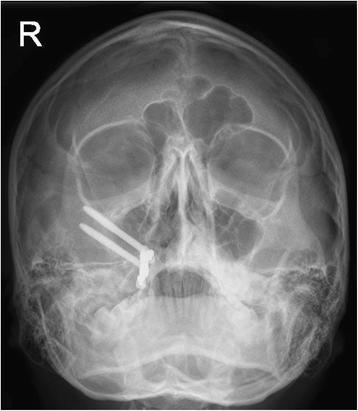
Fig. 11. Facial radiograph at 22-month follow-up : A novel report on the use of an oncology zygomatic implant
author: Amit Dattani, David Richardson, Chris J Butterworth | publisher: drg. Andreas Tjandra, Sp. Perio, FISID

Fig. 11. Facial radiograph at 22-month follow-up
Serial posts:
- Background : A novel report on the use of an oncology zygomatic implant-retained maxillary obturator in a paediatric patient [1]
- Background : A novel report on the use of an oncology zygomatic implant-retained maxillary obturator in a paediatric patient [2]
- Case presentation : A novel report on the use of an oncology zygomatic implant-retained maxillary obturator in a paediatric patient [1]
- Case presentation : A novel report on the use of an oncology zygomatic implant-retained maxillary obturator in a paediatric patient [2]
- Discussion : A novel report on the use of an oncology zygomatic implant-retained maxillary obturator in a paediatric patient [1]
- Discussion : A novel report on the use of an oncology zygomatic implant-retained maxillary obturator in a paediatric patient [2]
- Discussion : A novel report on the use of an oncology zygomatic implant-retained maxillary obturator in a paediatric patient [3]
- Conclusions : A novel report on the use of an oncology zygomatic implant-retained maxillary obturator in a paediatric patient
- References : A novel report on the use of an oncology zygomatic implant-retained maxillary obturator in a paediatric patient
- Author information : A novel report on the use of an oncology zygomatic implant-retained maxillary obturator in a paediatric patient
- Rights and permissions : A novel report on the use of an oncology zygomatic implant-retained maxillary obturator in a paediatric patient
- About this article : A novel report on the use of an oncology zygomatic implant-retained maxillary obturator in a paediatric patient
- Fig. 1. Zygomatic oncology implant with cleansable polished surface for intra-oral component : A novel report on the use of an oncology zygomatic implant
- Fig. 2. Palatal swelling (post-biopsy) between upper right first and second premolar teeth : A novel report on the use of an oncology zygomatic implant
- Fig. 3. Low-level right-sided maxillectomy with the insertion of two zygomatic oncology implants at time of surgery : A novel report on the use of an oncology zygomatic implant
- Fig. 4. Twelve-week review post-surgery prior to definitive impressions for the implant-supported prosthesis : A novel report on the use of an oncology zygomatic implant
- Fig. 5. Zygomatic implant bar utilising Rhein attachments for retention : A novel report on the use of an oncology zygomatic implant
- Fig. 6. Intaglio surface of definitive acrylic obturator with bar attachments in place. Note the absence of any other retaining clasps and the simple nature of this prosthesis : A novel report on the use of an oncology zygomatic implant
- Fig. 7. Smile view of definitive implant-retained obturator at initial fitting (April 2014) : A novel report on the use of an oncology zygomatic implant
- Fig. 8. Anterior view of definitive obturator prosthesis in occlusion : A novel report on the use of an oncology zygomatic implant
- Fig. 9. Palatal view of definitive implant-retained obturator at initial fitting (April 2014) : A novel report on the use of an oncology zygomatic implant
- Fig. 10. Full facial view of definitive implant-retained obturator at initial fitting (April 2014) : A novel report on the use of an oncology zygomatic implant
- Fig. 11. Facial radiograph at 22-month follow-up : A novel report on the use of an oncology zygomatic implant
- Fig. 12. Facial photograph views at 22-month follow-up : A novel report on the use of an oncology zygomatic implant