Panoramic radiography, Cone beam computed tomography, Maxillary sinus site, Subjective rating, Incidental radiographic findings, Education
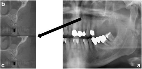
Fig. 2. a Panoramic radiography with area of interest (maxillary sinus) and b, c examples of corresponding images in cone beam computed tomography : Evaluation of symptomatic maxillary sinus patholog
author: Michael Dau, Paul Marciak, Bial Al-Nawas, Henning Staedt, Abdulmonem Alshiri, Bernhard Frerich, Peer Wolfgang Kmmerer | publisher: drg. Andreas Tjandra, Sp. Perio, FISID

Fig. 2. a Panoramic radiography with area of interest (maxillary sinus) and b, c examples of corresponding images in cone beam computed tomography
Serial posts:
- Background : Evaluation of symptomatic maxillary sinus pathologies using panoramic radiography and cone beam computed tomography—influence of professional training [1]
- Background : Evaluation of symptomatic maxillary sinus pathologies using panoramic radiography and cone beam computed tomography—influence of professional training [2]
- Methods : Evaluation of symptomatic maxillary sinus pathologies using panoramic radiography and cone beam computed tomography—influence of professional training [1]
- Methods : Evaluation of symptomatic maxillary sinus pathologies using panoramic radiography and cone beam computed tomography—influence of professional training [2]
- Results : Evaluation of symptomatic maxillary sinus pathologies using panoramic radiography and cone beam computed tomography—influence of professional training
- Discussion : Evaluation of symptomatic maxillary sinus pathologies using panoramic radiography and cone beam computed tomography—influence of professional training [1]
- Discussion : Evaluation of symptomatic maxillary sinus pathologies using panoramic radiography and cone beam computed tomography—influence of professional training [2]
- Conclusions : Evaluation of symptomatic maxillary sinus pathologies using panoramic radiography and cone beam computed tomography—influence of professional training
- References : Evaluation of symptomatic maxillary sinus pathologies using panoramic radiography and cone beam computed tomography—influence of professional training [1]
- References : Evaluation of symptomatic maxillary sinus pathologies using panoramic radiography and cone beam computed tomography—influence of professional training [2]
- References : Evaluation of symptomatic maxillary sinus pathologies using panoramic radiography and cone beam computed tomography—influence of professional training [3]
- References : Evaluation of symptomatic maxillary sinus pathologies using panoramic radiography and cone beam computed tomography—influence of professional training [4]
- Author information : Evaluation of symptomatic maxillary sinus pathologies using panoramic radiography and cone beam computed tomography—influence of professional training
- Rights and permissions : Evaluation of symptomatic maxillary sinus pathologies using panoramic radiography and cone beam computed tomography—influence of professional training
- About this article : Evaluation of symptomatic maxillary sinus pathologies using panoramic radiography and cone beam computed tomography—influence of professional training
- Table 1 Results of the question “Based on PAN, the clinical area of interest, is…” : Evaluation of symptomatic maxillary sinus pathologies using panoramic radiography and cone beam computed tomography—influence of professional training
- Table 2 Results of the question “An additional sFOV-CBCT of the clinical area of interest is…” : Evaluation of symptomatic maxillary sinus pathologies using panoramic radiography and cone beam computed tomography—influence of professional training
- Table 3 Number of additional incidental findings in PAN and sFOV-CBCT not related to the sinus disease that led to the radiographic examination : Evaluation of symptomatic maxillary sinus pathologies using panoramic radiography and cone beam computed tomography—influence of professional training
- Table 4 Description of incidental findings in PAN not related to the sinus disease that led to the radiographic examination : Evaluation of symptomatic maxillary sinus pathologies using panoramic radiography and cone beam computed tomography—influence of professional training
- Table 5 Results of the question “Is there an additional clinical value of sFOV-CBCT?” : Evaluation of symptomatic maxillary sinus pathologies using panoramic radiography and cone beam computed tomography—influence of professional training
- Fig. 1. a Panoramic radiography with area of interest (maxillary sinus) and b, c examples of corresponding images in cone beam computed tomography : Evaluation of symptomatic maxillary sinus patholog
- Fig. 2. a Panoramic radiography with area of interest (maxillary sinus) and b, c examples of corresponding images in cone beam computed tomography : Evaluation of symptomatic maxillary sinus patholog