Maxillary sinus endoscopy, Schneiderian membrane perforation, Crestal sinus lifter, Sinus implants, Endoscopic implants, Atrophic posterior maxilla
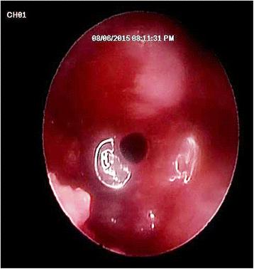
Fig. 5. Endoscopic view from the crestal osteotomy site showing perforation of the sinus lining under the power of magnification and illumination of the endoscope : Crestal endoscopic approach for evaluating sinus m
author: Samy Elian, Khaled Barakat | publisher: drg. Andreas Tjandra, Sp. Perio, FISID

Fig. 5. Endoscopic view from the crestal osteotomy site showing perforation of the sinus lining under the power of magnification and illumination of the endoscope
Serial posts:
- Introduction : Crestal endoscopic approach for evaluating sinus membrane elevation technique
- Patients and methods : Crestal endoscopic approach for evaluating sinus membrane elevation technique [1]
- Patients and methods : Crestal endoscopic approach for evaluating sinus membrane elevation technique [2]
- Results : Crestal endoscopic approach for evaluating sinus membrane elevation technique [1]
- Results : Crestal endoscopic approach for evaluating sinus membrane elevation technique [2]
- Discussion : Crestal endoscopic approach for evaluating sinus membrane elevation technique
- References : Crestal endoscopic approach for evaluating sinus membrane elevation technique [1]
- References : Crestal endoscopic approach for evaluating sinus membrane elevation technique [2]
- Acknowledgements : Crestal endoscopic approach for evaluating sinus membrane elevation technique
- Author information : Crestal endoscopic approach for evaluating sinus membrane elevation technique
- Ethics declarations : Crestal endoscopic approach for evaluating sinus membrane elevation technique
- Rights and permissions : Crestal endoscopic approach for evaluating sinus membrane elevation technique
- About this article : Crestal endoscopic approach for evaluating sinus membrane elevation technique
- Table 1 Descriptive statistics of membrane thickness and perforation rate : Crestal endoscopic approach for evaluating sinus membrane elevation technique
- Table 2 Chi square test showing perforation rate among different groups : Crestal endoscopic approach for evaluating sinus membrane elevation technique
- Table 3 Descriptive statistics, results of Kruskal-Wallis and Mann-Whitney U tests for comparison between membrane thicknesses of different morphologies : Crestal endoscopic approach for evaluating sinus membrane elevation technique
- Table 4 Chi square test showing perforation rate by different morphologies : Crestal endoscopic approach for evaluating sinus membrane elevation technique
- Fig. 1. A trephined hole (4 mm bone) in the lateral wall of the maxillary sinus to allow entrance of the endoscope : Crestal endoscopic approach for evaluating sinus m
- Fig. 2. Malleting instruments supplied from InnoBioSurg (IBS) Company, Korea. a magic sinus splitter: used to widen and split the crest. b magic sinus lifter: used to lift the available bone with its attached membrane : Crestal endoscopic approach for evaluating sinus m
- Fig. 3. Endoscopic view from the lateral sinus wall showing the dome-shape elevation of sinus lining : Crestal endoscopic approach for evaluating sinus m
- Fig. 4. Schematic drawing showing entrance of the endoscope from the crestal osteotomy site after sinus membrane elevation to assess the integrity of the membrane : Crestal endoscopic approach for evaluating sinus m
- Fig. 5. Endoscopic view from the crestal osteotomy site showing perforation of the sinus lining under the power of magnification and illumination of the endoscope : Crestal endoscopic approach for evaluating sinus m
- Fig. 6. Box plot representing mean values of membrane thicknesses for the investigated groups : Crestal endoscopic approach for evaluating sinus m
- Fig. 7. Box and Whisker plot representing median and range values of membrane thicknesses with different morphologies : Crestal endoscopic approach for evaluating sinus m