Fig. 6. Right sinus balloon dilation procedure. This photographic series shows the surgical procedure that augmented bone and allowed implant placement at the no. 3 site. a Preoperative view after infiltration anesthesia. b Full-thickness midcrestal incision. c Osteotomy preparation with implant drills and osteotomes. d, e The dilating balloon, which is inflated using saline pressure from a syringe. f Insertion of uninflated balloon into osteotomy. g Gentle inflation of balloon by 1 ml. h Preparation of allograft and collagen tape. i Collagen tape is visible at bottom of osteotomy after filling expanded Schneiderian membrane with bone graft and covering graft with collagen tape. j Implant placement. k Suturing with a continuous suture. l Postoperative radiograph showing implant and halo of allograft surrounding apex of implant after surgery : Case report on managing incomplete bone formation

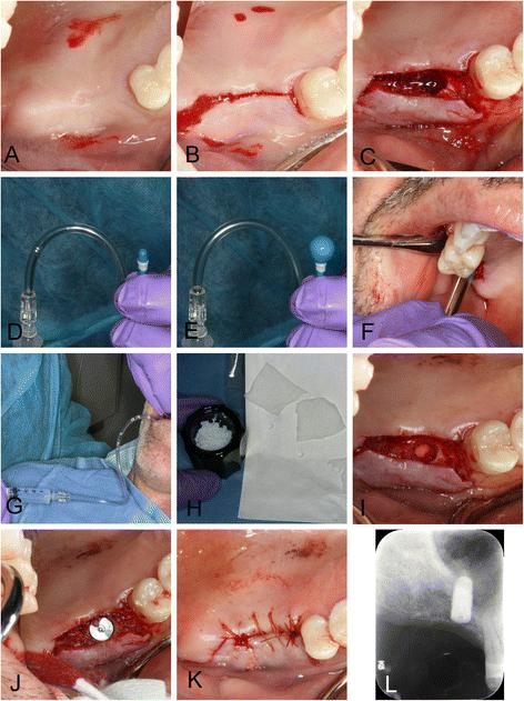
Fig. 6. Right sinus balloon dilation procedure. This photographic series shows the surgical procedure that augmented bone and allowed implant placement at the no. 3 site. a Preoperative view after infiltration anesthesia. b Full-thickness midcrestal incision. c Osteotomy preparation with implant drills and osteotomes. d, e The dilating balloon, which is inflated using saline pressure from a syringe. f Insertion of uninflated balloon into osteotomy. g Gentle inflation of balloon by 1 ml. h Preparation of allograft and collagen tape. i Collagen tape is visible at bottom of osteotomy after filling expanded Schneiderian membrane with bone graft and covering graft with collagen tape. j Implant placement. k Suturing with a continuous suture. l Postoperative radiograph showing implant and halo of allograft surrounding apex of implant after surgery
Serial posts:
- Background : Case report on managing incomplete bone formation after bilateral sinus augmentation using a palatal approach and a dilating balloon technique [1]
- Background : Case report on managing incomplete bone formation after bilateral sinus augmentation using a palatal approach and a dilating balloon technique [2]
- Case presentation : Case report on managing incomplete bone formation after bilateral sinus augmentation using a palatal approach and a dilating balloon technique [1]
- Case presentation : Case report on managing incomplete bone formation after bilateral sinus augmentation using a palatal approach and a dilating balloon technique [2]
- Case presentation : Case report on managing incomplete bone formation after bilateral sinus augmentation using a palatal approach and a dilating balloon technique [3]
- Case presentation : Case report on managing incomplete bone formation after bilateral sinus augmentation using a palatal approach and a dilating balloon technique [4]
- Case presentation : Case report on managing incomplete bone formation after bilateral sinus augmentation using a palatal approach and a dilating balloon technique [5]
- Conclusions : Case report on managing incomplete bone formation after bilateral sinus augmentation using a palatal approach and a dilating balloon technique
- References : Case report on managing incomplete bone formation after bilateral sinus augmentation using a palatal approach and a dilating balloon technique [1]
- References : Case report on managing incomplete bone formation after bilateral sinus augmentation using a palatal approach and a dilating balloon technique [2]
- Acknowledgements : Case report on managing incomplete bone formation after bilateral sinus augmentation using a palatal approach and a dilating balloon technique
- Author information : Case report on managing incomplete bone formation after bilateral sinus augmentation using a palatal approach and a dilating balloon technique
- Rights and permissions : Case report on managing incomplete bone formation after bilateral sinus augmentation using a palatal approach and a dilating balloon technique
- About this article : Case report on managing incomplete bone formation after bilateral sinus augmentation using a palatal approach and a dilating balloon technique
- Fig. 1. Initial presentation. Panoramic radiograph taken at initial visit shows severe bone loss, supraerupted molars and furcation involvement : Case report on managing incomplete bone formation
- Fig. 2. Right sinus prior to first sinus grafting procedure. Cone beam CT imaging shows very little residual bone volume at implant site for the no. 3 area : Case report on managing incomplete bone formation
- Fig. 3. Left sinus prior to first sinus grafting procedure. Cone beam CT imaging also shows very little bone volume on left side for the no. 14 area : Case report on managing incomplete bone formation
- Fig. 4. Right sinus about 12 months after first grafting procedure. Cone beam CT imaging shows little suitable bone at implant site, but grafted bone displaced distal to site. Bone hydroxyapatite particles were added as radiographic marker to the graft material for the first sinus augmentation procedure and are still visible as radiopaque specks : Case report on managing incomplete bone formation
- Fig. 5. Left sinus about 12 months after first grafting procedure. Cone beam CT imaging shows unusual sinus anatomy after grafting, with finger-like sinus extension at implant site, and thick-grafted bone buccal and apical to it. The infractured wall is still clearly visible, as well as the bovine bone particles used as radiographic marker : Case report on managing incomplete bone formation
- Fig. 6. Right sinus balloon dilation procedure. This photographic series shows the surgical procedure that augmented bone and allowed implant placement at the no. 3 site. a Preoperative view after infiltration anesthesia. b Full-thickness midcrestal incision. c Osteotomy preparation with implant drills and osteotomes. d, e The dilating balloon, which is inflated using saline pressure from a syringe. f Insertion of uninflated balloon into osteotomy. g Gentle inflation of balloon by 1 ml. h Preparation of allograft and collagen tape. i Collagen tape is visible at bottom of osteotomy after filling expanded Schneiderian membrane with bone graft and covering graft with collagen tape. j Implant placement. k Suturing with a continuous suture. l Postoperative radiograph showing implant and halo of allograft surrounding apex of implant after surgery : Case report on managing incomplete bone formation
- Fig. 7. Schematic diagram of sinus balloon dilating procedure. This diagram shows how the balloon is inserted into a small transcrestal osteotomy and then expanded with balloon : Case report on managing incomplete bone formation
- Fig. 8. Blood supply of the sinus. There are three areas in the sinus where blood vessels may be encountered during sinus augmentation procedures for implants. On the inflection point between hard palate and alveolar ridge in the posterior maxilla, the greater palatine neurovascular bundle is located embedded in soft tissue. This inflection point is matched in the internal sinus anatomy and presents a landmark that can be palpated with sinus curettes during sinus membrane elevation or seen on cone beam CT images in this patient. It is important to avoid instrumenting the area above this inflection point as branches of the lateral posterior nasal arteries may be encountered superior to this area. Injuring these blood vessels can lead to significant sinus bleeding that is difficult to stop without sinus tamponade. Often on cone beam CT images, we see a small blood vessel channel midway within the lateral wall of the sinus, which likely is the posterior superior alveolar artery and vein.
- Fig. 9. Schematic diagram of palatal approach sinus augmentation. The diagram shows the location of the lateral window, avoiding the thick grafted bone on the buccal, and the greater palatal neurovascular bundle : Case report on managing incomplete bone formation
- Fig. 10. Palatal approach lateral window sinus augmentation. This photographic series shows the surgical procedure that augmented bone and allowed implant placement at the no. 14 site. a Preoperative view prior to infiltration anesthesia. b Full-thickness midcrestal incision with palatal release and flap elevation. This was aided by a small bony ridge that separated the alveolar crest from the soft tissue area containing the greater palatine neurovascular bundle. c Sinus window created with piezosurgery. d–f With gentle piezocision and water pressure, the finger-like membrane is slowly mobilized and collapsed towards the remainder of the sinus cavity. The overlying bone serves to form a new floor covering the base of the finger-like cavity. f Conventional implant placement using osteotomy drills. g Any exposed sinus membrane is covered with collagen tape. h Particulate mineralized allograft is placed into the newly created space. i A resorbable collagen membrane is placed over the acce
- Fig. 11. Implant restoration. Implants were restored by dental students supervised by prosthodontists at the Dental Center : Case report on managing incomplete bone formation
- Fig. 12. Radiographic bone levels three years after placement. Bone levels remain unchanged during long-term follow-up : Case report on managing incomplete bone formation