Low-level maxillectomy, Zygomatic implants, Zygomatic oncology implant, Fixed dental prosthesis, ZIP flap, Micro-vascular reconstruction, Radiotherapy, Early implant loading, Oral cancer rehabilitation
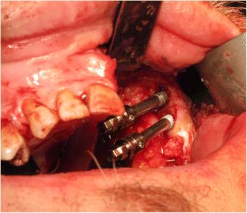
Fig. 6. Zygomatic oncology implants sited in the residual zygomatic bone on the defect side of the maxilla : The zygomatic implant
author: C J Butterworth, S N Rogers | publisher: drg. Andreas Tjandra, Sp. Perio, FISID

Fig. 6. Zygomatic oncology implants sited in the residual zygomatic bone on the defect side of the maxilla
Serial posts:
- Abstract : The zygomatic implant perforated (ZIP) flap: a new technique for combined surgical reconstruction and rapid fixed dental rehabilitation following low-level maxillectomy
- Background : The zygomatic implant perforated (ZIP) flap: a new technique for combined surgical reconstruction and rapid fixed dental rehabilitation following low-level maxillectomy
- Case presentation : The zygomatic implant perforated (ZIP) flap: a new technique for combined surgical reconstruction and rapid fixed dental rehabilitation following low-level maxillectomy [1]
- Case presentation : The zygomatic implant perforated (ZIP) flap: a new technique for combined surgical reconstruction and rapid fixed dental rehabilitation following low-level maxillectomy [2]
- Case presentation : The zygomatic implant perforated (ZIP) flap: a new technique for combined surgical reconstruction and rapid fixed dental rehabilitation following low-level maxillectomy [3]
- Case presentation : The zygomatic implant perforated (ZIP) flap: a new technique for combined surgical reconstruction and rapid fixed dental rehabilitation following low-level maxillectomy [4]
- Case presentation : The zygomatic implant perforated (ZIP) flap: a new technique for combined surgical reconstruction and rapid fixed dental rehabilitation following low-level maxillectomy [5]
- Case presentation : The zygomatic implant perforated (ZIP) flap: a new technique for combined surgical reconstruction and rapid fixed dental rehabilitation following low-level maxillectomy [6]
- Conclusions : The zygomatic implant perforated (ZIP) flap: a new technique for combined surgical reconstruction and rapid fixed dental rehabilitation following low-level maxillectomy
- References : The zygomatic implant perforated (ZIP) flap: a new technique for combined surgical reconstruction and rapid fixed dental rehabilitation following low-level maxillectomy
- Author information : The zygomatic implant perforated (ZIP) flap: a new technique for combined surgical reconstruction and rapid fixed dental rehabilitation following low-level maxillectomy
- Ethics declarations : The zygomatic implant perforated (ZIP) flap: a new technique for combined surgical reconstruction and rapid fixed dental rehabilitation following low-level maxillectomy
- Rights and permissions : The zygomatic implant perforated (ZIP) flap: a new technique for combined surgical reconstruction and rapid fixed dental rehabilitation following low-level maxillectomy
- About this article : The zygomatic implant perforated (ZIP) flap: a new technique for combined surgical reconstruction and rapid fixed dental rehabilitation following low-level maxillectomy
- Table 1 Patient-reported quality of life outcomes following ZIP flap procedure : The zygomatic implant perforated (ZIP) flap: a new technique for combined surgical reconstruction and rapid fixed dental rehabilitation following low-level maxillectomy
- Fig. 1. Clinical view of left-sided maxillary tumour at presentation : The zygomatic implant
- Fig. 2. Staging MRI scan showing destructive lesion left maxilla : The zygomatic implant
- Fig. 3. Staging CT scan confirming maxillary destruction but preservation of the orbital floor : The zygomatic implant
- Fig. 4. Panoramic dental radiograph showing dental status at presentation : The zygomatic implant
- Fig. 5. Left-sided maxillary resection (Brown class 2b) : The zygomatic implant
- Fig. 6. Zygomatic oncology implants sited in the residual zygomatic bone on the defect side of the maxilla : The zygomatic implant
- Fig. 7. Conventional zygomatic implant insertion on the non-defect side of the maxilla following extraction of the remaining teeth and an alveoloplasty : The zygomatic implant
- Fig. 8. Abutment level impression utilising light-cured acrylic tray material : The zygomatic implant
- Fig. 9. Inter-occlusal registration using the pre-fabricated maxillary denture prosthesis relined with silicone putty over the implant abutment protection caps : The zygomatic implant
- Fig. 10. Radial forearm flap inset and sutured into the maxillary defect and perforated by the zygomatic oncology implant abutments : The zygomatic implant
- Fig. 11. Intra-oral view of the soft tissue flap at 3 weeks post-operatively with overgrowth of flap over the zygomatic oncology implants : The zygomatic implant
- Fig. 12. Provisional acrylic fixed dental prosthesis fitted at 4 weeks post-surgery : The zygomatic implant
- Fig. 13. Panoramic dental radiograph showing the position of the zygomatic implants and the seating of the initial fixed prosthesis : The zygomatic implant
- Fig. 14. Intra-oral view of perforated flap 3 weeks following radiotherapy : The zygomatic implant
- Fig. 15. Facial appearance 18 months following treatment : The zygomatic implant
- Fig. 16. Another ZIP flap case demonstrating the use of a perforated polythene “washer” to keep the flap from overgrowing the implant abutments during the healing phase : The zygomatic implant
- Fig. 17. The appearance of the case shown in Fig. 16 with the polythene “washer” removed at 2 weeks post-surgery, providing access to the zygomatic oncology implants : The zygomatic implant