Oligodontia, Dental implants, Computer-guided implant dentistry, Guided surgery
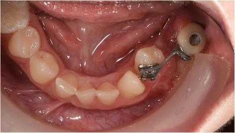
Fig. 8. Patient 2—intra-oral situation during orthodontic treatment at the age of 14. A temporary crown with bracket is fixed on the dental implant. Eight months after start of orthodontic treatment, the 34 is already close to the planned end position : Three-dimensional computer-guided implant
author: Marieke A P Filius, Joep Kraeima, Arjan Vissink, Krista I Janssen, Gerry M Raghoebar, Anita Visser | publisher: drg. Andreas Tjandra, Sp. Perio, FISID

Fig. 8. Patient 2—intra-oral situation during orthodontic treatment at the age of 14. A temporary crown with bracket is fixed on the dental implant. Eight months after start of orthodontic treatment, the 34 is already close to the planned end position
Serial posts:
- Abstract : Three-dimensional computer-guided implant placement in oligodontia
- Introduction : Three-dimensional computer-guided implant placement in oligodontia
- Patient and methods : Three-dimensional computer-guided implant placement in oligodontia [1]
- Patient and methods : Three-dimensional computer-guided implant placement in oligodontia [2]
- Results : Three-dimensional computer-guided implant placement in oligodontia
- Discussion : Three-dimensional computer-guided implant placement in oligodontia
- Conclusion : Three-dimensional computer-guided implant placement in oligodontia
- Abbreviations : Three-dimensional computer-guided implant placement in oligodontia
- References : Three-dimensional computer-guided implant placement in oligodontia
- Acknowledgements : Three-dimensional computer-guided implant placement in oligodontia
- Author information : Three-dimensional computer-guided implant placement in oligodontia [1]
- Author information : Three-dimensional computer-guided implant placement in oligodontia [2]
- Ethics declarations : Three-dimensional computer-guided implant placement in oligodontia
- Rights and permissions : Three-dimensional computer-guided implant placement in oligodontia
- About this article : Three-dimensional computer-guided implant placement in oligodontia
- Table 1 Accuracy data: Euclidian distances (ED, mm) of the apex (tip) and entry point (shoulder) and the degree (°) of angular deviation (axis) of the implants (n = 7) : Three-dimensional computer-guided implant placement in oligodontia
- Fig. 1. a Patient 1—orthopantomogram (OPT) at age of 13. Situation before extraction of the ankylosed deciduous teeth 55, 54, 65, 74, 75, 84, and 85 and start of orthodontic treatment. Eleven permanent teeth (including 4 third molars) were congenitally missing. b Patient 1—post-orthodontic situation at age of 16. The top of the mandibular processus alveolaris is small (upper). The interdental space at location of the second premolars in the maxilla is 7 and 14 mm at location of the premolars in the mandible. Six dental implants were planned (locations 15, 25, 34, 35, 44 and 45). Implant placement (inclusive bone augmentation with the autogenous retromolar mandibular bone 3 months before implant placement at the place of the 25) was postponed until the age of 18. Essix retainers were used to safeguard the width of the diastemas : Three-dimensional computer-guided implant
- Fig. 2. a Patient 2—pre-implant orthopantomogram (OPG) at the age of 12. Situation before start of orthodontic and implant treatment. Eleven permanent teeth (including 2 third molars) were congenitally missing and the 34 is impacted. To erect the 34, orthodontic treatment was desired. Due to the lack of stable anchorages in the third quadrant, it was decided to place one implant at tooth region 35 for orthodontic anchorage and future prosthetics. Due to very limited bone height virtual implant planning was needed to avoid damage to the mandibular nerve. b Patient 2—mandible, pre implant intra-oral situation at the age of 12. The 34 is not visible in the oral cavity : Three-dimensional computer-guided implant
- Fig. 3. a Patient 1—detailed 3D model of the combined data from the CBCT and intra-oral scan at age of 18. b Patient 2—detailed 3D model of the combined data from the CBCT and intra-oral scan at age of 12 : Three-dimensional computer-guided implant
- Fig. 4. a Patient 1—virtual set-up of the ultimate treatment goal. b Patient 2—virtual set-up of the ultimate implant position. One short dental implant was planned in region 35, based on the location of the mandibular nerve (orange), the impacted 34 (pink) and the bone quality and volume. c Patient 2—virtual set-up of the ultimate prosthetic treatment goal : Three-dimensional computer-guided implant
- Fig. 5. a Drilling templates of patient 1. Printed model of the maxilla (left) and mandible (right) with drilling template and metal drilling inserts (Nobel biocare). b Drilling template for the mandible of patient 1. c Implant placement of patient 1. Dental implant placement in the mandible using the virtual developed tooth-supported templates and metal drilling inserts : Three-dimensional computer-guided implant
- Fig. 6. Patient 1—post-operative orthopantomogram (OPT) at age of 18 : Three-dimensional computer-guided implant
- Fig. 7. Patient 2—post-operative orthopantomogram (OPT) at age of 13. Situation 10 months after implant placement. Three months after starting the orthodontic treatment, the 34 is already erected : Three-dimensional computer-guided implant
- Fig. 8. Patient 2—intra-oral situation during orthodontic treatment at the age of 14. A temporary crown with bracket is fixed on the dental implant. Eight months after start of orthodontic treatment, the 34 is already close to the planned end position : Three-dimensional computer-guided implant
- Fig. 9. Patient 1—prosthodontic end result 5 months after implant placement : Three-dimensional computer-guided implant
- Fig. 10. Patient 1—post-operative evaluation of placement accuracy of the implants in the mandible. Green is the planned position; blue is the actual position : Three-dimensional computer-guided implant