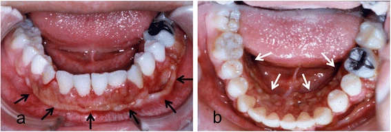
Figure 1. Intraoral photograph showing diffuse tumor formation on the alveolar gingiva (arrows)
Figure 1. Intraoral photograph
author: Kazuki Takaoka,Emi Segawa,Michiyo Yamamura,Yusuke Zushi,Masahiro UradeHiromitsu Kishimoto | publisher: drg. Andreas Tjandra, Sp. Perio, FISID

Serial posts:
- Dental implant treatment in a young woman
- Background : Dental implant treatment in a young woman
- Case presentation : Dental implant treatment in a young woman (1)
- Case presentation : Dental implant treatment in a young woman (2)
- Conclusion : Dental implant treatment in a young woman (1)
- Conclusion : Dental implant treatment in a young woman (2)
- Figure 1. Intraoral photograph
- Figure 2. Panoramic radiograph showing notable alveolar bone resorption
- Figure 4. Intraoperative photograph of resection of the alveolar ridge
- Figure 3. Photomicrographs of the biopsy specimen
- Figure 5. Preoperative intraoral photograph of implant placement
- Figure 6. a Mandibular implant-supported overdenture inserted into the mouth. b Panoramic radiograph after insertion of the prosthesis
- Figure 7. a Intraoral photograph. b Gold Dolder bar and screws; marked wear of a prosthetic screw (arrow)
- Figure 8. Periapical radiographs of the implants
- Figure 9. a Mandibular implant-fixed prosthesis inserted into the mouth.