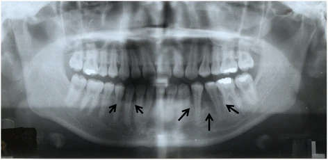
Figure 2. Panoramic radiograph showing notable alveolar bone resorption in the left mandibular premolar region and slight resorption in the right mandibular canine region (arrows)
Figure 2. Panoramic radiograph showing notable alveolar bone resorption
author: Kazuki Takaoka,Emi Segawa,Michiyo Yamamura,Yusuke Zushi,Masahiro UradeHiromitsu Kishimoto | publisher: drg. Andreas Tjandra, Sp. Perio, FISID

Serial posts:
- Dental implant treatment in a young woman
- Background : Dental implant treatment in a young woman
- Case presentation : Dental implant treatment in a young woman (1)
- Case presentation : Dental implant treatment in a young woman (2)
- Conclusion : Dental implant treatment in a young woman (1)
- Conclusion : Dental implant treatment in a young woman (2)
- Figure 1. Intraoral photograph
- Figure 2. Panoramic radiograph showing notable alveolar bone resorption
- Figure 4. Intraoperative photograph of resection of the alveolar ridge
- Figure 3. Photomicrographs of the biopsy specimen
- Figure 5. Preoperative intraoral photograph of implant placement
- Figure 6. a Mandibular implant-supported overdenture inserted into the mouth. b Panoramic radiograph after insertion of the prosthesis
- Figure 7. a Intraoral photograph. b Gold Dolder bar and screws; marked wear of a prosthetic screw (arrow)
- Figure 8. Periapical radiographs of the implants
- Figure 9. a Mandibular implant-fixed prosthesis inserted into the mouth.