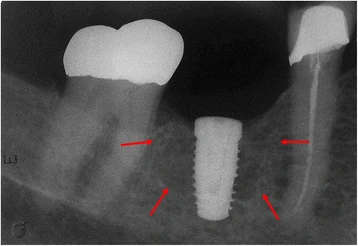
Figure 1. The periapical radiograph revealed the presence of an extensive and poorly circumscribed osteoporotic area around the proximal implant
Figure 1. The periapical radiograph revealed the presence of an extensive and poorly circumscribed osteoporotic area around the proximal implant
author: Natlia Galvo Garcia,Francisco Barbara Abreu Barros,Mrcia Maria Dalmolin Carvalho, Denise Tostes Oliveira | publisher: drg. Andreas Tjandra, Sp. Perio, FISID

Serial posts:
- Focal osteoporotic bone marrow defect involving dental implant (1)
- Focal osteoporotic bone marrow defect involving dental implant (2)
- Focal osteoporotic bone marrow defect involving dental implant (3)
- Figure 1. The periapical radiograph revealed the presence of an extensive and poorly circumscribed osteoporotic area around the proximal implant
- Figure 2. Normal hematopoietic cells, fat cells and bone trabeculae (hematoxylin and eosin, original magnification ×200)
- Figure 3. Erythroid, granulocytic, monocytic and lymphocytic series are illustrated, as well as megakaryocytes (hematoxylin and eosin, original magnification ×400)