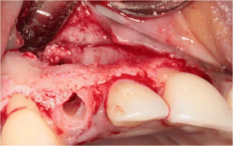
Figure 4. The prepared implant socket and osseous defect resulting from removal of the buccally impacted secondary canine and the primary canine. Note that the upper part of the alveolar crest is intact
Figure 4. The prepared implant socket and osseous defect
author: Elise G Zuiderveld,Henny J A Meijer,Arjan Vissink,Gerry M Raghoebar | publisher: drg. Andreas Tjandra, Sp. Perio, FISID

Serial posts:
- Immediate placement and provisionalization of an implant
- Background : Immediate placement and provisionalization of an implant
- Case presentation : Immediate placement and provisionalization of an implant (1)
- Case presentation : Immediate placement and provisionalization of an implant (2)
- Case presentation : Immediate placement and provisionalization of an implant (3)
- Case presentation : Immediate placement and provisionalization of an implant (4)
- Discussion : Immediate placement and provisionalization of an implant (4)
- Figure 1. Clinical view showing the failing right primary canine
- Figure 2. CBCT image showing the buccal location of the impacted secondary canine
- Figure 3. The impacted canine has become visible after elevation
- Figure 4. The prepared implant socket and osseous defect
- Figure 5. The implant is placed in the prepared socket
- Figure 6. Situation after implant placement and restoration
- Figure 7. Clinical view immediately after placement of the provisional implant crown
- Figure 8. The screw-retained definitive all-ceramic crown
- Figure 9. Intra-oral radiograph showing the implant 12 months after placement
- Figure 10. Clinical view showing the failing right primary canine
- Figure 11. CBCT image showing the palatal location of the impacted secondary canine
- Figure 12. The impacted canine has become visible
- Figure 13. Situation after implant placement and repair of the bony defect
- Figure 14. Clinical view showing optimal esthetics around the screw-retained definitive all-ceramic crown
- Figure 15. Intra-oral radiograph showing the implant 12 months after placement