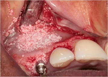
Figure 6. Situation after implant placement and restoration of the bony defect with a 1:1 mixture of Bio-Oss® and autologous bone
Figure 6. Situation after implant placement and restoration
author: Elise G Zuiderveld,Henny J A Meijer,Arjan Vissink,Gerry M Raghoebar | publisher: drg. Andreas Tjandra, Sp. Perio, FISID

Serial posts:
- Immediate placement and provisionalization of an implant
- Background : Immediate placement and provisionalization of an implant
- Case presentation : Immediate placement and provisionalization of an implant (1)
- Case presentation : Immediate placement and provisionalization of an implant (2)
- Case presentation : Immediate placement and provisionalization of an implant (3)
- Case presentation : Immediate placement and provisionalization of an implant (4)
- Discussion : Immediate placement and provisionalization of an implant (4)
- Figure 1. Clinical view showing the failing right primary canine
- Figure 2. CBCT image showing the buccal location of the impacted secondary canine
- Figure 3. The impacted canine has become visible after elevation
- Figure 4. The prepared implant socket and osseous defect
- Figure 5. The implant is placed in the prepared socket
- Figure 6. Situation after implant placement and restoration
- Figure 7. Clinical view immediately after placement of the provisional implant crown
- Figure 8. The screw-retained definitive all-ceramic crown
- Figure 9. Intra-oral radiograph showing the implant 12 months after placement
- Figure 10. Clinical view showing the failing right primary canine
- Figure 11. CBCT image showing the palatal location of the impacted secondary canine
- Figure 12. The impacted canine has become visible
- Figure 13. Situation after implant placement and repair of the bony defect
- Figure 14. Clinical view showing optimal esthetics around the screw-retained definitive all-ceramic crown
- Figure 15. Intra-oral radiograph showing the implant 12 months after placement