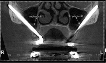
Figure 5. Coronal slice from the CBCT showing small exteriorization of a zygomatic implant apex
Figure 5. Coronal slice from the CBCT showing small exteriorization of a zygomatic implant apex
author: P P T Arajo, S A Sousa, V B S Diniz, P P Gomes, J S P da Silva, A R Germano | publisher: drg. Andreas Tjandra, Sp. Perio, FISID

Serial posts:
- Evaluation of patients undergoing placement of zygomatic implants using sinus slot technique
- Background : Evaluation of patients undergoing placement of zygomatic implants
- Methods : Evaluation of patients undergoing placement of zygomatic implants (1)
- Methods : Evaluation of patients undergoing placement of zygomatic implants (2)
- Methods : Evaluation of patients undergoing placement of zygomatic implants (3)
- Methods : Evaluation of patients undergoing placement of zygomatic implants (4)
- Methods : Evaluation of patients undergoing placement of zygomatic implants (5)
- Results : Evaluation of patients undergoing placement of zygomatic implants (1)
- Results : Evaluation of patients undergoing placement of zygomatic implants (2)
- Discussion : Evaluation of patients undergoing placement of zygomatic implants (1)
- Discussion : Evaluation of patients undergoing placement of zygomatic implants (2)
- Discussion : Evaluation of patients undergoing placement of zygomatic implants (3)
- Discussion : Evaluation of patients undergoing placement of zygomatic implants (4)
- Discussion : Evaluation of patients undergoing placement of zygomatic implants (5)
- Discussion : Evaluation of patients undergoing placement of zygomatic implants (6)
- Discussion : Evaluation of patients undergoing placement of zygomatic implants (7)
- Discussion : Evaluation of patients undergoing placement of zygomatic implants (8)
- Reference : Evaluation of patients undergoing placement of zygomatic implants (8)
- Figure 1. a Brånemark technique. b Sinus slot technique. c Extrasinus technique
- Figure 2. Periapical radiographs using the parallelism technique
- Figure 3. Panoramic radiograph showing bone level maintenance around the conventional implants
- Figure 4. Coronal slice from the CBCT showing implant apical third inside the zygomatic bone
- Figure 5. Coronal slice from the CBCT showing small exteriorization of a zygomatic implant apex
- Figure 6. Zygomatic implant probing using a WHO periodontal probe
- Figure 7. Visual analog scale—patient version
- Figure 8. Visual analog scale—evaluator version
- Table 1 Statistical analysis of individual parameters