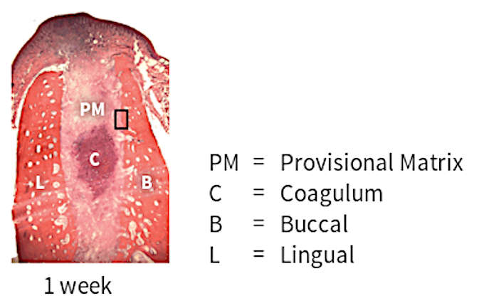
... the socket area is occupied by coagulum and granulation tissue.
Ridge alterations: 1 week
author: Nikos Mardas | publisher: drg. Andreas Tjandra, Sp. Perio, FISID

Araujo and Lindhe described the edentulous ridge profile alterations following tooth extraction in an experimental study in a dog model. During the first week of post-extraction healing, the socket area is occupied by coagulum and granulation tissue. A large number of osteoclasts are seen on the outer as well as on the inner surfaces of the buccal and lingual bone walls. The presence of osteoclasts on the inner surface of the socket walls indicates that the bundle bone resorption has been initiated.
Serial posts:
- Ridge alterations following tooth extraction
- Reduction in alveolar ridge characterizes alveolar atrophy
- Factors influence tissue atrophy
- Decrease in ridge height
- Decrease in ridge width
- Dimensional change in alveolar bone
- Mean width reduction
- Mean height reduction
- Radiographic height reduction
- Ridge alterations: 1 week
- Ridge alterations: 2 week
- Ridge alterations: 4 week
- Ridge alterations: 8 week
- Buccal wall
- Factors influencing post-extraction ridge atrophy