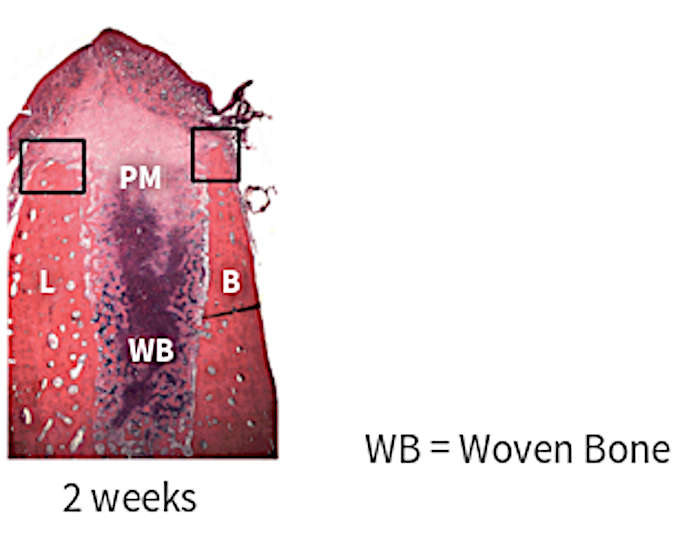
At 2, the apical and lateral parts of the socket are filled with woven bone
Ridge alterations: 2 week
author: Nikos Mardas | publisher: drg. Andreas Tjandra, Sp. Perio, FISID

At 2 weeks after tooth extraction, the apical and lateral parts of the socket are filled with woven bone, while the central and marginal portions of the socket are occupied by provisional connective tissue. On the inner and outer surfaces of the socket walls, numerous osteoclasts can be seen. In several areas of the socket wall, the bundle bone has been resorbed and replaced with woven bone.
Serial posts:
- Ridge alterations following tooth extraction
- Reduction in alveolar ridge characterizes alveolar atrophy
- Factors influence tissue atrophy
- Decrease in ridge height
- Decrease in ridge width
- Dimensional change in alveolar bone
- Mean width reduction
- Mean height reduction
- Radiographic height reduction
- Ridge alterations: 1 week
- Ridge alterations: 2 week
- Ridge alterations: 4 week
- Ridge alterations: 8 week
- Buccal wall
- Factors influencing post-extraction ridge atrophy