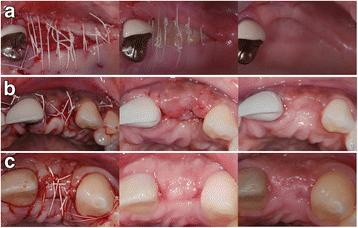
Fig. 1. Clinical photographs of the both treatment groups after the initial surgery, 1 week post-op and at the re-entry. a) In the test group, no primary wound closure was achieved (left) and the barrier was left exposed for secondary intention healing. After 1 week, the matrix remained exposed (middle) showing no signs of infection. For months later, the exposed area was covered by a keratinized tissue (right). b) In the test group, primary wound closure was achieved at surgery (left). However, the barrier became exposed after 1 week of healing (middle). For months later, exposed area was covered with a keratinized tissue (right). c) In the control group, primary wound closure was achieved (left). After 1 week (middle), primary healing happened without any signs of membrane exposure. For months later, the site healed uneventfully (right) : The effect of membrane exposure on lateral ridge a

Fig. 1. Clinical photographs of the both treatment groups after the initial surgery, 1 week post-op and at the re-entry. a) In the test group, no primary wound closure was achieved (left) and the barrier was left exposed for secondary intention healing. After 1 week, the matrix remained exposed (middle) showing no signs of infection. For months later, the exposed area was covered by a keratinized tissue (right). b) In the test group, primary wound closure was achieved at surgery (left). However, the barrier became exposed after 1 week of healing (middle). For months later, exposed area was covered with a keratinized tissue (right). c) In the control group, primary wound closure was achieved (left). After 1 week (middle), primary healing happened without any signs of membrane exposure. For months later, the site healed uneventfully (right)
Serial posts:
- Abstract : The effect of membrane exposure on lateral ridge augmentation: a case-controlled study
- Background : The effect of membrane exposure on lateral ridge augmentation: a case-controlled study [1]
- Methods : The effect of membrane exposure on lateral ridge augmentation: a case-controlled study [1]
- Methods : The effect of membrane exposure on lateral ridge augmentation: a case-controlled study [2]
- Results : The effect of membrane exposure on lateral ridge augmentation: a case-controlled study
- Discussion : The effect of membrane exposure on lateral ridge augmentation: a case-controlled study [1]
- Discussion : The effect of membrane exposure on lateral ridge augmentation: a case-controlled study [2]
- Conclusions : The effect of membrane exposure on lateral ridge augmentation: a case-controlled study
- References : The effect of membrane exposure on lateral ridge augmentation: a case-controlled study [1]
- References : The effect of membrane exposure on lateral ridge augmentation: a case-controlled study [2]
- References : The effect of membrane exposure on lateral ridge augmentation: a case-controlled study [3]
- References : The effect of membrane exposure on lateral ridge augmentation: a case-controlled study [4]
- Acknowledgements : The effect of membrane exposure on lateral ridge augmentation: a case-controlled study
- Author information : The effect of membrane exposure on lateral ridge augmentation: a case-controlled study
- Rights and permissions : The effect of membrane exposure on lateral ridge augmentation: a case-controlled study
- About this article : The effect of membrane exposure on lateral ridge augmentation: a case-controlled study
- Table 1 Patient population and demographics and sites : The effect of membrane exposure on lateral ridge augmentation: a case-controlled study
- Table 2 Baseline and re-entry measurement of the alveolar ridge width : The effect of membrane exposure on lateral ridge augmentation: a case-controlled study
- Table 3 Alveolar ridge width reduction : The effect of membrane exposure on lateral ridge augmentation: a case-controlled study
- Fig. 1. Clinical photographs of the both treatment groups after the initial surgery, 1 week post-op and at the re-entry. a) In the test group, no primary wound closure was achieved (left) and the barrier was left exposed for secondary intention healing. After 1 week, the matrix remained exposed (middle) showing no signs of infection. For months later, the exposed area was covered by a keratinized tissue (right). b) In the test group, primary wound closure was achieved at surgery (left). However, the barrier became exposed after 1 week of healing (middle). For months later, exposed area was covered with a keratinized tissue (right). c) In the control group, primary wound closure was achieved (left). After 1 week (middle), primary healing happened without any signs of membrane exposure. For months later, the site healed uneventfully (right) : The effect of membrane exposure on lateral ridge a