Bone atrophy, Bone resorption, Dental implants, Implant failure, Narrow-diameter implants, Posterior mandible
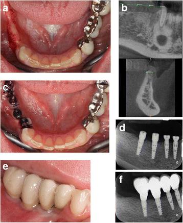
Fig. 2. Case 1: Example of one case involved in the study. a Preoperative view of a partial edentulism in posterior mandible. b Preoperative CT scan. The width of the ridge was 4 mm. c Four narrow diameter implants were placed and left to a nonsubmerged healing. d Baseline periapical radiograph. e Buccal vieew of the final metal ceramic restoration. f Periapical radiograph at 1 year after loading : Narrow implant
author: Tommaso Grandi, Luigi Svezia, Giovanni Grandi | publisher: drg. Andreas Tjandra, Sp. Perio, FISID

Fig. 2. Case 1: Example of one case involved in the study. a Preoperative view of a partial edentulism in posterior mandible. b Preoperative CT scan. The width of the ridge was 4 mm. c Four narrow diameter implants were placed and left to a nonsubmerged healing. d Baseline periapical radiograph. e Buccal vieew of the final metal ceramic restoration. f Periapical radiograph at 1 year after loading
Serial posts:
- Abstract : Narrow implants (2.75 and 3.25 mm diameter) supporting a fixed splinted prostheses in posterior regions of mandible: one-year results from a prospective cohort study
- Background : Narrow implants (2.75 and 3.25 mm diameter) supporting a fixed splinted prostheses in posterior regions of mandible: one-year results from a prospective cohort study [1]
- Background : Narrow implants (2.75 and 3.25 mm diameter) supporting a fixed splinted prostheses in posterior regions of mandible: one-year results from a prospective cohort study [2]
- Methods : Narrow implants (2.75 and 3.25 mm diameter) supporting a fixed splinted prostheses in posterior regions of mandible: one-year results from a prospective cohort study [1]
- Methods : Narrow implants (2.75 and 3.25 mm diameter) supporting a fixed splinted prostheses in posterior regions of mandible: one-year results from a prospective cohort study [2]
- Results : Narrow implants (2.75 and 3.25 mm diameter) supporting a fixed splinted prostheses in posterior regions of mandible: one-year results from a prospective cohort study [1]
- Results : Narrow implants (2.75 and 3.25 mm diameter) supporting a fixed splinted prostheses in posterior regions of mandible: one-year results from a prospective cohort study [2]
- Discussion : Narrow implants (2.75 and 3.25 mm diameter) supporting a fixed splinted prostheses in posterior regions of mandible: one-year results from a prospective cohort study [1]
- Discussion : Narrow implants (2.75 and 3.25 mm diameter) supporting a fixed splinted prostheses in posterior regions of mandible: one-year results from a prospective cohort study [2]
- Conclusions : Narrow implants (2.75 and 3.25 mm diameter) supporting a fixed splinted prostheses in posterior regions of mandible: one-year results from a prospective cohort study
- References : Narrow implants (2.75 and 3.25 mm diameter) supporting a fixed splinted prostheses in posterior regions of mandible: one-year results from a prospective cohort study [1]
- References : Narrow implants (2.75 and 3.25 mm diameter) supporting a fixed splinted prostheses in posterior regions of mandible: one-year results from a prospective cohort study [2]
- Author information : Narrow implants (2.75 and 3.25 mm diameter) supporting a fixed splinted prostheses in posterior regions of mandible: one-year results from a prospective cohort study
- Ethics declarations : Narrow implants (2.75 and 3.25 mm diameter) supporting a fixed splinted prostheses in posterior regions of mandible: one-year results from a prospective cohort study
- Rights and permissions : Narrow implants (2.75 and 3.25 mm diameter) supporting a fixed splinted prostheses in posterior regions of mandible: one-year results from a prospective cohort study
- About this article : Narrow implants (2.75 and 3.25 mm diameter) supporting a fixed splinted prostheses in posterior regions of mandible: one-year results from a prospective cohort study
- Table 1 Features of the subjects included in the study : Narrow implants (2.75 and 3.25 mm diameter) supporting a fixed splinted prostheses in posterior regions of mandible: one-year results from a prospective cohort study
- Table 2 Dimensions (diameter and length) and final seating torque of the inserted implants (n = 124) : Narrow implants (2.75 and 3.25 mm diameter) supporting a fixed splinted prostheses in posterior regions of mandible: one-year results from a prospective cohort study
- Table 3 Comparison of mean bone levels (means ± SD) at different follow-up intervals : Narrow implants (2.75 and 3.25 mm diameter) supporting a fixed splinted prostheses in posterior regions of mandible: one-year results from a prospective cohort study
- Table 4 Comparison of mean bone levels (means ± SD) at different follow-up intervals in different implants diameters groups (2.75 and 3.25 mm) : Narrow implants (2.75 and 3.25 mm diameter) supporting a fixed splinted prostheses in posterior regions of mandible: one-year results from a prospective cohort study
- Fig. 1. Characteristics of the implants used in the study: a external macro-design of JDIcon Ultra S, 2.75 mm diameter implant and b external macro-design of JDEvolution S, 3.25 mm diameter implant : Narrow implant
- Fig. 2. Case 1: Example of one case involved in the study. a Preoperative view of a partial edentulism in posterior mandible. b Preoperative CT scan. The width of the ridge was 4 mm. c Four narrow diameter implants were placed and left to a nonsubmerged healing. d Baseline periapical radiograph. e Buccal vieew of the final metal ceramic restoration. f Periapical radiograph at 1 year after loading : Narrow implant
- Fig. 3. Example of another case involved in the study. a Preoperative view –premolars and molars are missing in left mandible. b Preoperative CT scan. The width of the ridge was around 4 mm. c Baseline periapical radiograph. Four narrow diameter implants were placed to restore the area. d Buccal view of the final full-contour zirconia restoration. e Periapical radiograph at 1 year after loading : Narrow implant