Explantation, Counter/torque, Dental implant, Periimplantitis, Implant removal, Minimally invasive
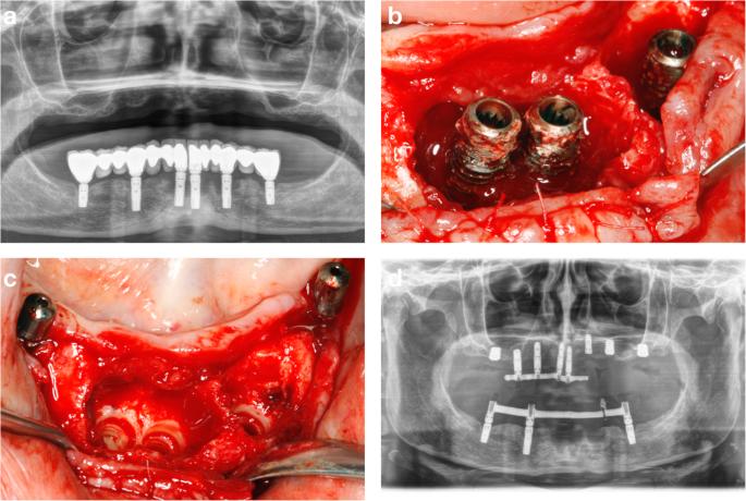
Fig. 3. Panoramic radiograph showing excessive marginal bone loss affecting all the dental implants in the mandible supporting fixed prostheses (a). Clinical image showing the advanced bone destruction around the implants at the incisors and left premolar regions (b). Clinical image showing the preservation of the pre-existing bone upon implant removal with the counter-torque regions (c). Panoramic radiograph showing the maintenance at this stage of 3 implants to support the provisional prosthesis in the mandible (d) : Performance of the counter-torque technique in the explantation of nonmobile dental implant
author: Eduardo Anitua, Sofia Fernandez-de-Retana, Mohammad H Alkhraisat | publisher: drg. Andreas Tjandra, Sp. Perio, FISID

Fig. 3. Panoramic radiograph showing excessive marginal bone loss affecting all the dental implants in the mandible supporting fixed prostheses (a). Clinical image showing the advanced bone destruction around the implants at the incisors and left premolar regions (b). Clinical image showing the preservation of the pre-existing bone upon implant removal with the counter-torque regions (c). Panoramic radiograph showing the maintenance at this stage of 3 implants to support the provisional prosthesis in the mandible (d)
Serial posts:
- Abstract : Performance of the counter-torque technique in the explantation of nonmobile dental implants
- Background : Performance of the counter-torque technique in the explantation of nonmobile dental implants
- Methods : Performance of the counter-torque technique in the explantation of nonmobile dental implants
- Results : Performance of the counter-torque technique in the explantation of nonmobile dental implants [1]
- Results : Performance of the counter-torque technique in the explantation of nonmobile dental implants [2]
- Discussion : Performance of the counter-torque technique in the explantation of nonmobile dental implants
- Conclusions : Performance of the counter-torque technique in the explantation of nonmobile dental implants
- Availability of data and materials : Performance of the counter-torque technique in the explantation of nonmobile dental implants
- References : Performance of the counter-torque technique in the explantation of nonmobile dental implants
- Acknowledgements : Performance of the counter-torque technique in the explantation of nonmobile dental implants
- Funding : Performance of the counter-torque technique in the explantation of nonmobile dental implants
- Author information : Performance of the counter-torque technique in the explantation of nonmobile dental implants
- Ethics declarations : Performance of the counter-torque technique in the explantation of nonmobile dental implants
- Additional information : Performance of the counter-torque technique in the explantation of nonmobile dental implants
- Rights and permissions : Performance of the counter-torque technique in the explantation of nonmobile dental implants
- About this article : Performance of the counter-torque technique in the explantation of nonmobile dental implants
- Table 1 Description of the implants fractured during the explantation : Performance of the counter-torque technique in the explantation of nonmobile dental implants
- Fig. 1. Frequency distribution of the location of the explanted dental implant : Performance of the counter-torque technique in the explantation of nonmobile dental implant
- Fig. 2. The cause of implant removal : Performance of the counter-torque technique in the explantation of nonmobile dental implant
- Fig. 3. Panoramic radiograph showing excessive marginal bone loss affecting all the dental implants in the mandible supporting fixed prostheses (a). Clinical image showing the advanced bone destruction around the implants at the incisors and left premolar regions (b). Clinical image showing the preservation of the pre-existing bone upon implant removal with the counter-torque regions (c). Panoramic radiograph showing the maintenance at this stage of 3 implants to support the provisional prosthesis in the mandible (d) : Performance of the counter-torque technique in the explantation of nonmobile dental implant
- Fig. 4. Implant placement surgery after 4 months of healing (a). Immediate loading of the new implants and the explanation of the implant at the left first molar (b). Panoramic radiograph showing the case finished with 12 months of follow-up (c). Clinical image showing the definitive screw-retained prostheses (d) : Performance of the counter-torque technique in the explantation of nonmobile dental implant