Animal study, Sinus floor elevation, Bone window, Bone, Bone plate, Polylactic membrane, Window repositioning, Bone graft
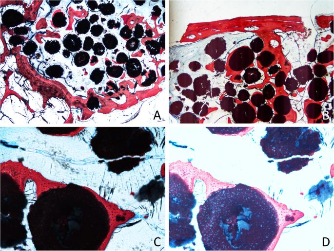
Fig. 4. Photomicrographs of ground sections after 4 months of healing. a Bone formed from the base of the sinus. b Bone plate connected by bridges of the new bone to the close-to-window region. c Particle of the graft surrounded by new bone. d Overexposed image to show the new bone ingrowth within the granules of biomaterial : Bone plate repositioned over the antrostomy after
author: Alessandro Perini, Giada Ferrante, Stefano Sivolella, Joaqun Urbizo Velez, Franco Bengazi, Daniele Botticelli | publisher: drg. Andreas Tjandra, Sp. Perio, FISID

Fig. 4. Photomicrographs of ground sections after 4 months of healing. a Bone formed from the base of the sinus. b Bone plate connected by bridges of the new bone to the close-to-window region. c Particle of the graft surrounded by new bone. d Overexposed image to show the new bone ingrowth within the granules of biomaterial
Serial posts:
- Abstract : Bone plate repositioned over the antrostomy after sinus floor elevation: an experimental study in sheep
- Introduction : Bone plate repositioned over the antrostomy after sinus floor elevation: an experimental study in sheep [1]
- Introduction : Bone plate repositioned over the antrostomy after sinus floor elevation: an experimental study in sheep [2]
- Materials and methods : Bone plate repositioned over the antrostomy after sinus floor elevation: an experimental study in sheep [1]
- Materials and methods : Bone plate repositioned over the antrostomy after sinus floor elevation: an experimental study in sheep [2]
- Materials and methods : Bone plate repositioned over the antrostomy after sinus floor elevation: an experimental study in sheep [3]
- Materials and methods : Bone plate repositioned over the antrostomy after sinus floor elevation: an experimental study in sheep [4]
- Results : Bone plate repositioned over the antrostomy after sinus floor elevation: an experimental study in sheep [1]
- Results : Bone plate repositioned over the antrostomy after sinus floor elevation: an experimental study in sheep [2]
- Discussion : Bone plate repositioned over the antrostomy after sinus floor elevation: an experimental study in sheep [1]
- Discussion : Bone plate repositioned over the antrostomy after sinus floor elevation: an experimental study in sheep [2]
- Discussion : Bone plate repositioned over the antrostomy after sinus floor elevation: an experimental study in sheep [3]
- Conclusion : Bone plate repositioned over the antrostomy after sinus floor elevation: an experimental study in sheep
- Availability of data and materials : Bone plate repositioned over the antrostomy after sinus floor elevation: an experimental study in sheep
- Abbreviations : Bone plate repositioned over the antrostomy after sinus floor elevation: an experimental study in sheep
- References : Bone plate repositioned over the antrostomy after sinus floor elevation: an experimental study in sheep [1]
- References : Bone plate repositioned over the antrostomy after sinus floor elevation: an experimental study in sheep [2]
- References : Bone plate repositioned over the antrostomy after sinus floor elevation: an experimental study in sheep [3]
- References : Bone plate repositioned over the antrostomy after sinus floor elevation: an experimental study in sheep [4]
- Acknowledgements : Bone plate repositioned over the antrostomy after sinus floor elevation: an experimental study in sheep
- Funding : Bone plate repositioned over the antrostomy after sinus floor elevation: an experimental study in sheep
- Author information : Bone plate repositioned over the antrostomy after sinus floor elevation: an experimental study in sheep [1]
- Author information : Bone plate repositioned over the antrostomy after sinus floor elevation: an experimental study in sheep [2]
- Ethics declarations : Bone plate repositioned over the antrostomy after sinus floor elevation: an experimental study in sheep
- Additional information : Bone plate repositioned over the antrostomy after sinus floor elevation: an experimental study in sheep
- Rights and permissions : Bone plate repositioned over the antrostomy after sinus floor elevation: an experimental study in sheep
- About this article : Bone plate repositioned over the antrostomy after sinus floor elevation: an experimental study in sheep
- Table 1 Percentages of the various tissues within the elevated area after 4 months of healing. Mean values ± standard deviations (P values) and median (25%; 75% percentiles) : Bone plate repositioned over the antrostomy after sinus floor elevation: an experimental study in sheep
- Table 2 Percentages of various tissues in the antrostomy area after 4 months of healing. Mean values ± standard deviations (P values) and median (25%; 75% percentiles) : Bone plate repositioned over the antrostomy after sinus floor elevation: an experimental study in sheep
- Fig. 1. Clinical view at a membrane site. a Skin and periosteum were separately elevated, and the facial sinus wall exposed. b A 12 × 8-mm window was cut and removed. c The Schneiderian membrane was carefully elevated. d A twisted wire was inserted in the middle of the long side of the window and the elevated sinus was grafted. e At the control site, a resorbable membrane was placed and secured with cyanoacrylate. f Membrane in situ : Bone plate repositioned over the antrostomy after
- Fig. 2. Clinical view at a bone plate site. a The bone window was removed. b The sinus mucosa was carefully elevated, and a twisted wire was placed. c The elevated sinus was grafted. d The access bony window was repositioned and secured with cyanoacrylate : Bone plate repositioned over the antrostomy after
- Fig. 3. a The elevated area was divided into four regions for morphometric analysis. RED: submucosa; GREEN: middle; YELLOW: base; PURPLE: close-to-window. INC: top of the infraorbital nerve canal : Bone plate repositioned over the antrostomy after
- Fig. 4. Photomicrographs of ground sections after 4 months of healing. a Bone formed from the base of the sinus. b Bone plate connected by bridges of the new bone to the close-to-window region. c Particle of the graft surrounded by new bone. d Overexposed image to show the new bone ingrowth within the granules of biomaterial : Bone plate repositioned over the antrostomy after
- Fig. 5. Graph representing the tissue percentages within the elevated area. No statistically significant differences were found : Bone plate repositioned over the antrostomy after
- Fig. 6. Graph representing new bone and composite bone percentages within the elevated area : Bone plate repositioned over the antrostomy after