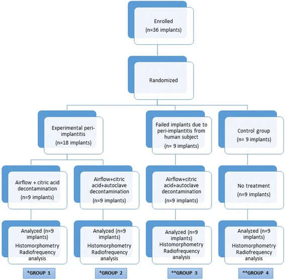
Figure 1. Flowchart of the research design employed in the study. *Three dogs were used in each group 1 and 2. Three implants were inserted right side of the mandibles. After peri-implantitis period, extracted implants were inserted into the left side of the mandibles. **Two dogs were used in each group 3 and 4. Six failed implants from human inserted into the one dog’s mandible bilaterally and three implants inserted into the other dog’s mandible unilaterally in group 3. Six implants inserted into the one dog’s mandible bilaterally and three implants inserted into the other dog’s mandible unilaterally in group 4 (control group)
Figure 1. Flowchart of the research design employed in the study
author: Murat Ulu,Erdem Kl,Emrah Soylu,Mehmet Krk, Alper Alkan | publisher: drg. Andreas Tjandra, Sp. Perio, FISID

Serial posts:
- Reusing dental implants
- Background : Reusing dental implants
- Methods : Reusing dental implants (1)
- Methods : Reusing dental implants (2)
- Methods : Reusing dental implants (3)
- Methods : Reusing dental implants (4)
- Results : Reusing dental implants
- Discussion : Reusing dental implants (1)
- Discussion : Reusing dental implants (2)
- Discussion : Reusing dental implants (3)
- Table 1 Comparison of BIC percentages of over the entire implant length at 3-month follow-up
- Table 2 Comparison of BIC percentages of 3 mm crestal area of the implants
- Table 3 Inter- and intra-group ISQ analysis and measurements on day of surgery and at 3-month follow-up
- Figure 1. Flowchart of the research design employed in the study
- Figure 2. Edentulous posterior mandible of the dog at 3 months after tooth extraction
- Figure 3. Silk ligatures placed in a submarginal position around the implants
- Figure 4. A 2-month period was allowed for plaque retention and peri-implantitis
- Figure 5. Time arrow about the stages of the study
- Figure 6. BIC percentage measured with ImageJ analysis software