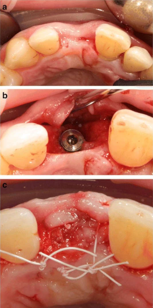
Figure 2. Clinical image of patient 4: a region 21 before implant placement. b, c Implant placement using the GBR procedure with a synthetic bone substitute material composed of HA + β-TCP
Figure 2. Clinical image of patient 4
author: Jonas Lorenz,Henriette Lerner, Robert A Sader, Shahram Ghanaati | publisher: drg. Andreas Tjandra, Sp. Perio, FISID

Serial posts:
- Investigation of peri-implant in implants
- Background: Investigation of peri-implant in implants (1)
- Background: Investigation of peri-implant in implants (2)
- Background: Investigation of peri-implant in implants (3)
- Methods: Investigation of peri-implant in implants (1)
- Methods: Investigation of peri-implant in implants (2)
- Results: Investigation of peri-implant in implants
- Discussion: Investigation of peri-implant in implants (1)
- Discussion: Investigation of peri-implant in implants (2)
- Discussion: Investigation of peri-implant in implants (3)
- Discussion: Investigation of peri-implant in implants
- Table 1 Participating patients and the number and site of the inserted implants
- Table 2 Results from the clinical and radiological 3-year follow-up investigation
- Figure 1. Schematic representation of the technical characteristics
- Figure 2. Clinical image of patient 4