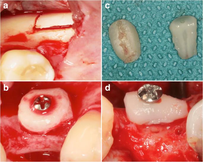
Fig. 1. Lateral ridge augmentation—a surgical procedure in the AB and TR groups

Fig. 1. Lateral ridge augmentation—a surgical procedure in the AB and TR groups. a The retromolar area served as a donor site for the harvesting of monocortical bone blocks in the AB group. b AB blocks were shaped to match the size and configuration of the defect site and fixed using one central osteosynthesis screw. c TR grafts were separated from either partially/fully retained or impacted wisdom teeth. d The most suitable specimen was positioned and fixed in a way that the exposed dentin faced the defect area, thus facilitating ankylosis at the recipient site. The crestal perforations were derived from initial attempts to pre-drill the osteosynthesis screw. All sites were left to heal in a submerged position without providing any contour augmentation procedures
Serial posts:
- Abstract : Radiographic outcomes following lateral alveolar ridge augmentation using autogenous tooth roots
- Background : Radiographic outcomes following lateral alveolar ridge augmentation using autogenous tooth roots
- Methods : Radiographic outcomes following lateral alveolar ridge augmentation using autogenous tooth roots [1]
- Methods : Radiographic outcomes following lateral alveolar ridge augmentation using autogenous tooth roots [2]
- Methods : Radiographic outcomes following lateral alveolar ridge augmentation using autogenous tooth roots [3]
- Results : Radiographic outcomes following lateral alveolar ridge augmentation using autogenous tooth roots
- Discussion : Radiographic outcomes following lateral alveolar ridge augmentation using autogenous tooth roots [1]
- Discussion : Radiographic outcomes following lateral alveolar ridge augmentation using autogenous tooth roots [2]
- Conclusions : Radiographic outcomes following lateral alveolar ridge augmentation using autogenous tooth roots
- References : Radiographic outcomes following lateral alveolar ridge augmentation using autogenous tooth roots
- Author information : Radiographic outcomes following lateral alveolar ridge augmentation using autogenous tooth roots
- Ethics declarations : Radiographic outcomes following lateral alveolar ridge augmentation using autogenous tooth roots
- Rights and permissions : Radiographic outcomes following lateral alveolar ridge augmentation using autogenous tooth roots
- About this article : Radiographic outcomes following lateral alveolar ridge augmentation using autogenous tooth roots
- Table 1 Study design and follow up visits : Radiographic outcomes following lateral alveolar ridge augmentation using autogenous tooth roots
- Table 2 Secondary performance endpoints (in mm) : Radiographic outcomes following lateral alveolar ridge augmentation using autogenous tooth roots
- Fig. 1. Lateral ridge augmentation—a surgical procedure in the AB and TR groups
- Fig. 2. Radiographic assessments. Images of the coronal planes representing the most central aspect of the respective defect sites were analyzed for the basal graft integration (i.e., contact between the graft and the host bone in %) (BI26) and the cross-sectional grafted area (mm2) (SA26) : Radiographic outcomes following lateral alveolar r
- Fig. 3. Representative CBCT outcomes at 26 weeks. a, b TR graft. c, d AB graft : Radiographic outcomes following lateral alveolar r
- Fig. 4. Linear regression plots to depict the relationship between BI26 and CWb/SA26 values. a CWb (TR group). b CWb (AB group). c SA26 (TR group). d SA26 (AB group) : Radiographic outcomes following lateral alveolar r