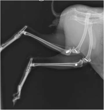
Figure 1. Radiograph showing implants in the rabbit tibia
Figure 1. Radiograph showing implants in the rabbit tibia
author: Jagjit S Dhaliwal,Rubens F Albuquerque Jr,Monzur MurshedJocelyne S Feine | publisher: drg. Andreas Tjandra, Sp. Perio, FISID

Serial posts:
- Osseointegration of standard and mini dental implants: a histomorphometric comparison
- Background : Osseointegration of standard and mini dental implants (1)
- Background : Osseointegration of standard and mini dental implants (2)
- Methods : Osseointegration of standard and mini dental implants (1)
- Methods : Osseointegration of standard and mini dental implants (2)
- Methods : Osseointegration of standard and mini dental implants (3)
- Methods : Osseointegration of standard and mini dental implants (4)
- Methods : Osseointegration of standard and mini dental implants (5)
- Methods : Osseointegration of standard and mini dental implants (6)
- Results : Osseointegration of standard and mini dental implants
- Discussion : Osseointegration of standard and mini dental implants (1)
- Discussion : Osseointegration of standard and mini dental implants (2)
- Figure 1. Radiograph showing implants in the rabbit tibia
- Figure 2. Leica SP 1600 saw microtome
- Figure 3. Histological sections being obtained with Leica SP 1600 saw microtome
- Figure 4. Histological section of mini dental implant in rabbit tibia stained with methylene blue and basic fuchsin
- Figure 5. Histological section of standard implant in rabbit tibia stained with methylene blue and basic fuchsin
- Figure 6. Micro CT scan images of the MDIs and Ankylos® embedded in rabbit bone 6 weeks post implantation
- Table 1 Comparison of % BIC in both groups
- Table 2 Descriptive statistics of the experimental and control group