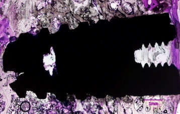
Figure 5. Histological section of standard implant in rabbit tibia stained with methylene blue and basic fuchsin
Figure 5. Histological section of standard implant in rabbit tibia stained with methylene blue and basic fuchsin
author: Jagjit S Dhaliwal,Rubens F Albuquerque Jr,Monzur MurshedJocelyne S Feine | publisher: drg. Andreas Tjandra, Sp. Perio, FISID

Serial posts:
- Osseointegration of standard and mini dental implants: a histomorphometric comparison
- Background : Osseointegration of standard and mini dental implants (1)
- Background : Osseointegration of standard and mini dental implants (2)
- Methods : Osseointegration of standard and mini dental implants (1)
- Methods : Osseointegration of standard and mini dental implants (2)
- Methods : Osseointegration of standard and mini dental implants (3)
- Methods : Osseointegration of standard and mini dental implants (4)
- Methods : Osseointegration of standard and mini dental implants (5)
- Methods : Osseointegration of standard and mini dental implants (6)
- Results : Osseointegration of standard and mini dental implants
- Discussion : Osseointegration of standard and mini dental implants (1)
- Discussion : Osseointegration of standard and mini dental implants (2)
- Figure 1. Radiograph showing implants in the rabbit tibia
- Figure 2. Leica SP 1600 saw microtome
- Figure 3. Histological sections being obtained with Leica SP 1600 saw microtome
- Figure 4. Histological section of mini dental implant in rabbit tibia stained with methylene blue and basic fuchsin
- Figure 5. Histological section of standard implant in rabbit tibia stained with methylene blue and basic fuchsin
- Figure 6. Micro CT scan images of the MDIs and Ankylos® embedded in rabbit bone 6 weeks post implantation
- Table 1 Comparison of % BIC in both groups
- Table 2 Descriptive statistics of the experimental and control group