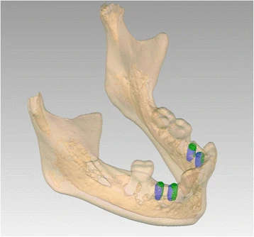
Figure 10. Patient 1—post-operative evaluation of placement accuracy of the implants in the mandible. Green is the planned position; blue is the actual position
Figure 10. Patient 1—post-operative evaluation of placement accuracy of the implants
author: Marieke A P Filius,Joep Kraeima,Arjan Vissink,Krista I Janssen,Gerry M Raghoebar,Anita Visser | publisher: drg. Andreas Tjandra, Sp. Perio, FISID

Serial posts:
- Three-dimensional computer-guided implant placement in oligodontia
- Introduction : Three-dimensional computer-guided implant placement in oligodontia
- Methods : Three-dimensional computer-guided implant placement in oligodontia
- Results : Three-dimensional computer-guided implant placement in oligodontia
- Figure 1. Patient 1—orthopantomogram (OPT) at age of 13
- Figure 2 a Patient 2—pre-implant orthopantomogram
- Figure 3. a Patient 1—detailed 3D model of the combined data
- Figure 4. a Patient 1—virtual set-up of the ultimate treatment goal
- Figure 5. a Drilling templates of patient 1
- Figure 6. Patient 1—post-operative orthopantomogram (OPT) at age of 18
- Figure 7. Patient 2—post-operative orthopantomogram (OPT) at age of 13. Situation 10 months after implant placement. Three months after starting the orthodontic treatment, the 34 is already erected
- Figure 8. Patient 2—intra-oral situation during orthodontic treatment
- Figure 9. Patient 1—prosthodontic end result 5 months after implant placement
- Figure 10. Patient 1—post-operative evaluation of placement accuracy of the implants