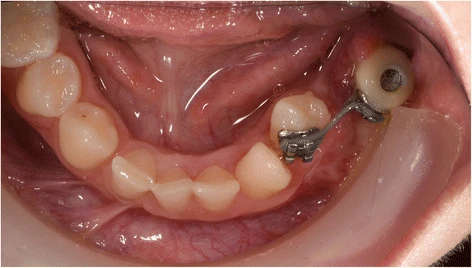
Figure 8. Patient 2—intra-oral situation during orthodontic treatment at the age of 14. A temporary crown with bracket is fixed on the dental implant. Eight months after start of orthodontic treatment, the 34 is already close to the planned end position
Figure 8. Patient 2—intra-oral situation during orthodontic treatment
author: Marieke A P Filius,Joep Kraeima,Arjan Vissink,Krista I Janssen,Gerry M Raghoebar,Anita Visser | publisher: drg. Andreas Tjandra, Sp. Perio, FISID

Serial posts:
- Three-dimensional computer-guided implant placement in oligodontia
- Introduction : Three-dimensional computer-guided implant placement in oligodontia
- Methods : Three-dimensional computer-guided implant placement in oligodontia
- Results : Three-dimensional computer-guided implant placement in oligodontia
- Figure 1. Patient 1—orthopantomogram (OPT) at age of 13
- Figure 2 a Patient 2—pre-implant orthopantomogram
- Figure 3. a Patient 1—detailed 3D model of the combined data
- Figure 4. a Patient 1—virtual set-up of the ultimate treatment goal
- Figure 5. a Drilling templates of patient 1
- Figure 6. Patient 1—post-operative orthopantomogram (OPT) at age of 18
- Figure 7. Patient 2—post-operative orthopantomogram (OPT) at age of 13. Situation 10 months after implant placement. Three months after starting the orthodontic treatment, the 34 is already erected
- Figure 8. Patient 2—intra-oral situation during orthodontic treatment
- Figure 9. Patient 1—prosthodontic end result 5 months after implant placement
- Figure 10. Patient 1—post-operative evaluation of placement accuracy of the implants