Many reports of the relationship between dental implants and osteoporosis have been published, but few have focused on bone quality.
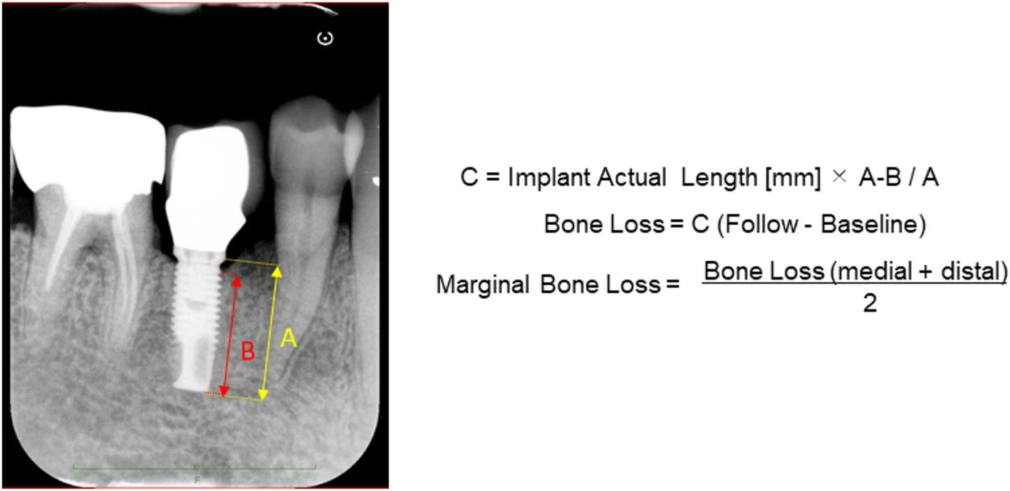
Figure 2. Measurement of marginal bone loss (MBL) on dental radiography.
author: Keisuke Yasuda,Shinsuke Okada,Yohei Okazaki,Kyou Hiasa,Kazuhiro Tsuga, Yasuhiko Abe | publisher: drg. Andreas Tjandra, Sp. Perio, FISID

Serial posts:
- Bone turnover markers to assess jawbone quality prior to dental implant treatment: a case-control study
- Background : Bone turnover markers to assess jawbone quality prior to dental implant treatment
- Background : Bone turnover markers to assess jawbone quality prior to dental implant treatment (1)
- Background : Bone turnover markers to assess jawbone quality prior to dental implant treatment (2)
- Materials and methods : Bone turnover markers to assess jawbone quality prior to dental implant treatment (1)
- Results : Bone turnover markers to assess jawbone quality prior to dental implant treatment (2)
- Discussion : Bone turnover markers to assess jawbone quality prior to dental implant treatment (1)
- Discussion : Bone turnover markers to assess jawbone quality prior to dental implant treatment (2)
- Discussion : Bone turnover markers to assess jawbone quality prior to dental implant treatment (3)
- Figure 1. Measure the bone density at the implant placement sites
- Figure 2. Measurement of marginal bone loss (MBL) on dental radiography.
- Table 1 Each parameter of the 18 patients who fulfilled the inclusion criteria
- Table 2 Age, sex, and follow-up period between the normal and abnormal group
- Figure 3. The overview on BTM values are shown
- Figure 4. Cancellous bone densities in the normal and abnormal groups of women
- Figure 5. Cancellous bone densities in SLA and MK-III implants
- Figure 6. Marginal bone loss (MBL) in SLA and MK-III implants