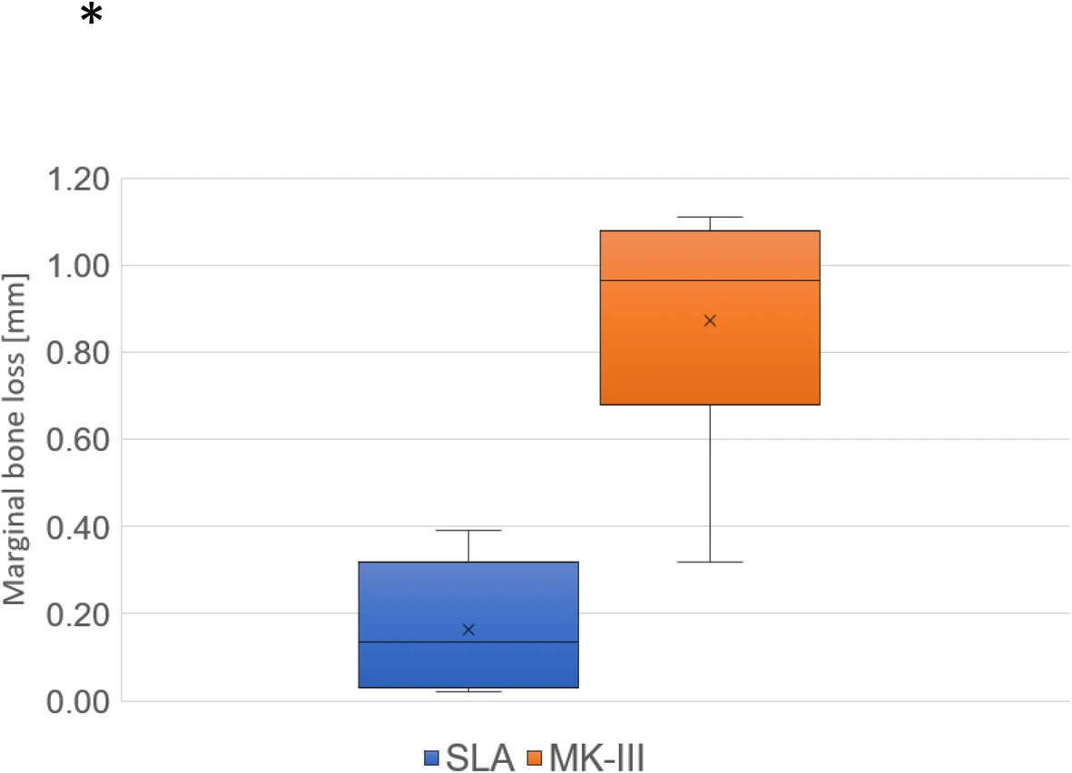
Figure 6. Marginal bone loss (MBL) in SLA and MK-III implants within the abnormal group. MBL is significantly larger in MK-III implants than SLA implants during the follow-up period.
Figure 6. Marginal bone loss (MBL) in SLA and MK-III implants
author: Keisuke Yasuda,Shinsuke Okada,Yohei Okazaki,Kyou Hiasa,Kazuhiro Tsuga, Yasuhiko Abe | publisher: drg. Andreas Tjandra, Sp. Perio, FISID

Serial posts:
- Bone turnover markers to assess jawbone quality prior to dental implant treatment: a case-control study
- Background : Bone turnover markers to assess jawbone quality prior to dental implant treatment
- Background : Bone turnover markers to assess jawbone quality prior to dental implant treatment (1)
- Background : Bone turnover markers to assess jawbone quality prior to dental implant treatment (2)
- Materials and methods : Bone turnover markers to assess jawbone quality prior to dental implant treatment (1)
- Results : Bone turnover markers to assess jawbone quality prior to dental implant treatment (2)
- Discussion : Bone turnover markers to assess jawbone quality prior to dental implant treatment (1)
- Discussion : Bone turnover markers to assess jawbone quality prior to dental implant treatment (2)
- Discussion : Bone turnover markers to assess jawbone quality prior to dental implant treatment (3)
- Figure 1. Measure the bone density at the implant placement sites
- Figure 2. Measurement of marginal bone loss (MBL) on dental radiography.
- Table 1 Each parameter of the 18 patients who fulfilled the inclusion criteria
- Table 2 Age, sex, and follow-up period between the normal and abnormal group
- Figure 3. The overview on BTM values are shown
- Figure 4. Cancellous bone densities in the normal and abnormal groups of women
- Figure 5. Cancellous bone densities in SLA and MK-III implants
- Figure 6. Marginal bone loss (MBL) in SLA and MK-III implants