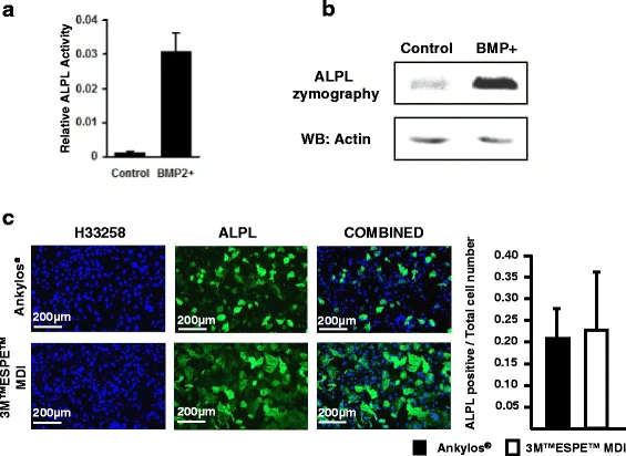
Figure 4. a C2C12 cells (control) and pBMP-2-transfected C2C12 cells were seeded on a 24-well plate (50,000 cell/well) and cultured in DMEM medium for 48 h. ALPL assay showing upregulated ALPL activity in the BMP-2-transfected C2C12 cells. b Cell extracts of C2C12 cells and pBMP-2-transfected cells were run on a 10% SDS-PAGE under non-denaturing conditions. The gel was then stained with NBT/BCIP solution (upper panel). Western bloting of actin showing the equal protein loading on the gel (lower panel). c Increased proliferation of C2C12 cells transfected with BMP-2 as well as ALPL activity when seeded on 3M™ESPE™ MDI disks. However, when the number of ALPL-positive cells is normalized to the total cell number, no differences are observed
Figure 4. a C2C12 cells (control) and pBMP-2-transfected C2C12 cells
author: Jagjit Singh Dhaliwal,Juliana Marulanda,Jingjing Li,Sharifa Alebrahim,Jocelyne Sheila Feine, Monzur Murshed | publisher: drg. Andreas Tjandra, Sp. Perio, FISID

Serial posts:
- In vitro comparison of two titanium dental implant surface treatments
- Background : Comparison of two titanium dental implant surface treatments
- Methods : Comparison of two titanium dental implant surface treatments (1)
- Results : Comparison of two titanium dental implant surface treatments
- Methods : Comparison of two titanium dental implant surface treatments (2)
- Methods : Comparison of two titanium dental implant surface treatments (3)
- Discussion : Comparison of two titanium dental implant surface treatments (1)
- Discussion : Comparison of two titanium dental implant surface treatments (2)
- Discussion : Comparison of two titanium dental implant surface treatments (3)
- Figure 1. Preparation of specimens
- Figure 2. Implant surface characterization under SEM
- Figure 3. Increased proliferation of C2C12 cells grown
- Figure 4. a C2C12 cells (control) and pBMP-2-transfected C2C12 cells
- Figure 5. Florescence microscopy