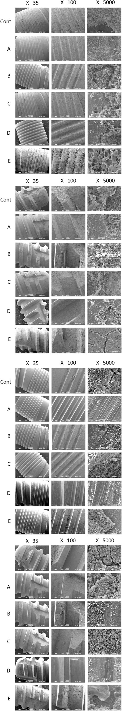
Figure 4. SEM analysis of 4 areas. 1 Rough surface—microthread area. 2 Rough surface—macrothread area. 3 Machined surface—microthread area. 4 Machined surface—macrothread area
Figure 4. SEM analysis of 4 areas. 1 Rough surface—microthread area
author: Motohiro Otsuki, Masahiro Wada, Masaya Yamaguchi, Shigetada Kawabata, Yoshinobu Maeda Kazunori Ikebe | publisher: drg. Andreas Tjandra, Sp. Perio, FISID
Figure 4. SEM analysis of 4 areas. 1 Rough surface—microthread area. 2 Rough surface—macrothread area. 3 Machined surface—microthread area. 4 Machined surface—macrothread area
Serial posts:
-
Evaluation of decontamination methods of oral biofilms formed on screw-shaped, rough and machined surface implants: an ex vivo study
-
Background : Evaluation of decontamination methods of oral biofilms formed on screw-shaped, rough and machined surface implants
-
Materials & methods : Evaluation of decontamination methods on implants (1)
-
Materials & methods : Evaluation of decontamination methods on implants (2)
-
Materials & methods : Evaluation of decontamination methods on implants (3)
-
Results : Evaluation of decontamination methods on implants (3)
-
Discussion : Evaluation of decontamination methods on implants (1)
-
Discussion : Evaluation of decontamination methods on implants (2)
-
Discussion : Evaluation of decontamination methods on implants (3)
-
Discussion : Evaluation of decontamination methods on implants (4)
-
Discussion : Evaluation of decontamination methods on implants (5)
-
Discussion : Evaluation of decontamination methods on implants (6)
-
Discussion : Evaluation of decontamination methods on implants (7)
-
Discussion : Evaluation of decontamination methods on implants (8)
-
Discussion : Evaluation of decontamination methods on implants (9)
-
Figure 1. Hard resin splint model carrying 6 implants
-
Figure 2. GC Aadva® implant; 3.3-mm diameter, 8-mm length
-
Figure 3. Decontamination methods
-
Figure 4. SEM analysis of 4 areas. 1 Rough surface—microthread area
-
Figure 5. Quantitative analysis of CFU counts on implants
-
Figure 6. Comparison of cleansability of each decontamination method
-
Table 1 Qualitative evaluation by SEM analysis of micro- and macrothread areas of rough surface implants
-
Table 2 Qualitative evaluation by SEM analysis of micro- and macrothread areas of machined surface implants
-
Table 3 Quantitative analysis of CFU counts
Figure 4. SEM analysis of 4 areas. 1 Rough surface—microthread area. 2 Rough surface—macrothread area. 3 Machined surface—microthread area. 4 Machined surface—macrothread area
- Evaluation of decontamination methods of oral biofilms formed on screw-shaped, rough and machined surface implants: an ex vivo study
- Background : Evaluation of decontamination methods of oral biofilms formed on screw-shaped, rough and machined surface implants
- Materials & methods : Evaluation of decontamination methods on implants (1)
- Materials & methods : Evaluation of decontamination methods on implants (2)
- Materials & methods : Evaluation of decontamination methods on implants (3)
- Results : Evaluation of decontamination methods on implants (3)
- Discussion : Evaluation of decontamination methods on implants (1)
- Discussion : Evaluation of decontamination methods on implants (2)
- Discussion : Evaluation of decontamination methods on implants (3)
- Discussion : Evaluation of decontamination methods on implants (4)
- Discussion : Evaluation of decontamination methods on implants (5)
- Discussion : Evaluation of decontamination methods on implants (6)
- Discussion : Evaluation of decontamination methods on implants (7)
- Discussion : Evaluation of decontamination methods on implants (8)
- Discussion : Evaluation of decontamination methods on implants (9)
- Figure 1. Hard resin splint model carrying 6 implants
- Figure 2. GC Aadva® implant; 3.3-mm diameter, 8-mm length
- Figure 3. Decontamination methods
- Figure 4. SEM analysis of 4 areas. 1 Rough surface—microthread area
- Figure 5. Quantitative analysis of CFU counts on implants
- Figure 6. Comparison of cleansability of each decontamination method
- Table 1 Qualitative evaluation by SEM analysis of micro- and macrothread areas of rough surface implants
- Table 2 Qualitative evaluation by SEM analysis of micro- and macrothread areas of machined surface implants
- Table 3 Quantitative analysis of CFU counts