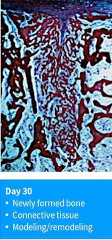
At 30 days of healing, a significant part of the extraction socket is filled with newly formed bone.
Day 30 - 60: Modeling/ remodeling
author: Nikos Mardas | publisher: drg. Andreas Tjandra, Sp. Perio, FISID

At 30 days of healing, a significant part of the extraction socket is filled with newly formed bone. This bone contains a large number of primary osteons and is continuous with the original bone of the socket walls. In some areas, the process of modeling and remodeling of the newly formed bone has begun. Osteoclasts are present on the surface of the original cortical bone lateral to the crestal region of the extraction socket.
Serial posts:
- Biological events after tooth extraction
- Day 1 : blood coagulation
- Day 3 : tissue composition in extraction socket
- Day 3 : At the end of this initial healing period
- Day 7: Provisional matrix & osteoclasts
- Day 14 : Woven bone & connective tissue
- Day 30 - 60: Modeling/ remodeling
- Day 120: corticular bone & trabecular bone
- Day 180: large marrow spaces
- Quantitative tissue analysis
- Post molar extraction mandible