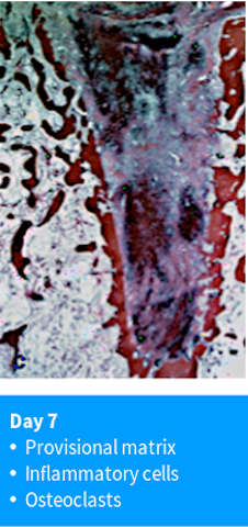
Day 7: Provisional matrix & osteoclasts

After 1 week of healing, the wound in the extraction site has significantly changed. In the central and apical part of the socket, large areas of the coagulum have been replaced with a provisional connective tissue matrix, which is lightly stained in the histologic section. Regions of darker-staining granulation tissue can still be seen. This provisional matrix is made of newly formed connective tissue fibers, blood vessels, mesenchymal cells, and various types of leukocytes. At the margins of the socket, the bone has begun to lose its continuity due to the action of osteoclasts, which have started to resorb the bundle bone. The gaps in the bone that connect the inner part of the socket to the surrounding trabecular bone are also known as Volkmann's canals. These gaps allow new blood vessels to grow into the socket from the surrounding bone.
Serial posts:
- Biological events after tooth extraction
- Day 1 : blood coagulation
- Day 3 : tissue composition in extraction socket
- Day 3 : At the end of this initial healing period
- Day 7: Provisional matrix & osteoclasts
- Day 14 : Woven bone & connective tissue
- Day 30 - 60: Modeling/ remodeling
- Day 120: corticular bone & trabecular bone
- Day 180: large marrow spaces
- Quantitative tissue analysis
- Post molar extraction mandible