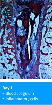
Hard and soft tissue composition in the extraction socket during longer healing periods was documented
Day 1 : blood coagulation
author: Nikos Mardas | publisher: drg. Andreas Tjandra, Sp. Perio, FISID

Hard and soft tissue composition in the extraction socket during longer healing periods was documented in a study by Cardaropoli and coworkers, in which the entire healing cascade after tooth extraction was evaluated in a dog model. The following images are histologic specimens representing the stages of healing observed in their study. On the day of extraction, the outline of the socket can be seen as a region of pink-stained bone. The inner lining of the bone is bundle bone, which was previously attached to the extracted tooth by the periodontal ligament. Within the socket, the coagulum appears as a non-homogenous mixture of fibrin, red blood cells, and inflammatory cells.
Serial posts:
- Biological events after tooth extraction
- Day 1 : blood coagulation
- Day 3 : tissue composition in extraction socket
- Day 3 : At the end of this initial healing period
- Day 7: Provisional matrix & osteoclasts
- Day 14 : Woven bone & connective tissue
- Day 30 - 60: Modeling/ remodeling
- Day 120: corticular bone & trabecular bone
- Day 180: large marrow spaces
- Quantitative tissue analysis
- Post molar extraction mandible