Cone-beam computed tomography, CBCT, Digital imaging, Maxillary sinus, Volumetric analysis
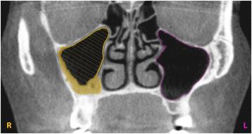
Fig. 2. View from the coronal plane. The marked curves define the osseus and mucous boundaries of the maxillary sinuses. The hatched surface illustrates the measured remaining pneumatized cavity of an obliterated sinus and the filled (yellow) surface highlights the calculated obliterated volume : 3D-evaluation of the maxillary sinus in cone-beam
author: Julia Luz, Dominique Greutmann, Daniel Wiedemeier, Claudio Rostetter, Martin Rcker, Bernd Stadlinger | publisher: drg. Andreas Tjandra, Sp. Perio, FISID

Fig. 2. View from the coronal plane. The marked curves define the osseus and mucous boundaries of the maxillary sinuses. The hatched surface illustrates the measured remaining pneumatized cavity of an obliterated sinus and the filled (yellow) surface highlights the calculated obliterated volume
Serial posts:
- Abstract : 3D-evaluation of the maxillary sinus in cone-beam computed tomography
- Background : 3D-evaluation of the maxillary sinus in cone-beam computed tomography
- Methods : 3D-evaluation of the maxillary sinus in cone-beam computed tomography [1]
- Methods : 3D-evaluation of the maxillary sinus in cone-beam computed tomography [2]
- Results : 3D-evaluation of the maxillary sinus in cone-beam computed tomography [1]
- Results : 3D-evaluation of the maxillary sinus in cone-beam computed tomography [2]
- Discussion : 3D-evaluation of the maxillary sinus in cone-beam computed tomography [1]
- Discussion : 3D-evaluation of the maxillary sinus in cone-beam computed tomography [2]
- Conclusions : 3D-evaluation of the maxillary sinus in cone-beam computed tomography
- References : 3D-evaluation of the maxillary sinus in cone-beam computed tomography [1]
- References : 3D-evaluation of the maxillary sinus in cone-beam computed tomography [2]
- References : 3D-evaluation of the maxillary sinus in cone-beam computed tomography [3]
- Author information : 3D-evaluation of the maxillary sinus in cone-beam computed tomography [1]
- Author information : 3D-evaluation of the maxillary sinus in cone-beam computed tomography [2]
- Ethics declarations : 3D-evaluation of the maxillary sinus in cone-beam computed tomography
- Rights and permissions : 3D-evaluation of the maxillary sinus in cone-beam computed tomography
- About this article : 3D-evaluation of the maxillary sinus in cone-beam computed tomography
- Table 1 Mean, median minimum, maximum, and standard deviation of the surface in square centimeter and volume in cubic centimeter of the osseus maxillary sinuses and the remaining pneumatized cavities in cases of obliterated sinuses as well as mean, median, minimum, maximum, and standard deviation of the calculated obliterated sinus volume in cubic centimeter : 3D-evaluation of the maxillary sinus in cone-beam computed tomography
- Table 2 Frequency of pathologies in 128 maxillary sinuses : 3D-evaluation of the maxillary sinus in cone-beam computed tomography
- Fig. 1. Calculation of the sinus body by interpolating 15–25 curves at a distance of 2 mm, depending upon the size of the maxillary cavity : 3D-evaluation of the maxillary sinus in cone-beam
- Fig. 2. View from the coronal plane. The marked curves define the osseus and mucous boundaries of the maxillary sinuses. The hatched surface illustrates the measured remaining pneumatized cavity of an obliterated sinus and the filled (yellow) surface highlights the calculated obliterated volume : 3D-evaluation of the maxillary sinus in cone-beam
- Fig. 3. 3D view of osseus sinus volumes. Surface area (cm2) and volume (cm3) were calculated by the software : 3D-evaluation of the maxillary sinus in cone-beam
- Fig. 4. The association between the osseus volume and the dentition. Edentulous, partly edentulous, and dentate patients showed no relevant difference in the size of the osseus sinus volume (p = 0.52) : 3D-evaluation of the maxillary sinus in cone-beam
- Fig. 5. The association between the obliterated volume and sinus pathology. The presence of a pathology significantly increased the obliterated volume of a maxillary sinus (p < 0.001). For better visibility, the diagram has been jittered along the x-axis : 3D-evaluation of the maxillary sinus in cone-beam
- Fig. 6. No statistical significant association between a patient’s age and the presence of obliteration of at least one maxillary sinus was found (p = 0.92). For better visibility, the diagram has been jittered along the y-axis : 3D-evaluation of the maxillary sinus in cone-beam
- Fig. 7. The association between the mean osseus sinus volume and age. No significant association between these parameters was found (p = 0.2) : 3D-evaluation of the maxillary sinus in cone-beam
- Fig. 8. The association between mean osseus sinus volume of the maxillary sinus and gender. Men have a statistically significant higher mean osseus volume than women (p = 0.007) : 3D-evaluation of the maxillary sinus in cone-beam