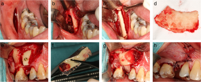
Figure 1. Intraoperative photos illustrating bone harvesting and lateral bone augmentation in the PRF group. Initially, an incision is made at the lateral aspect of the posterior part of the mandibular corpus (a) followed by exposing the mucoperiosteal flap (b), before making the osteotomy line (c). The bone block (d) is then retrieved before adjusted to the contour at the recipient site and fixated with an osteosynthesis screw (e). Autogenous bone graft particles are packed around the graft before covering the grafted area with three PRF membranes (f and g). Finally, tension-free primary wound closure is performed before suturing (h)
Figure 1. Intraoperative photos illustrating bone harvesting
author: Jens Hartlev,Sren Schou,Flemming Isidor, Sven Erik Nrholt | publisher: drg. Andreas Tjandra, Sp. Perio, FISID

Serial posts:
- A clinical and radiographic study of implants placed in autogenous bone grafts
- Background: A clinical and radiographic study of implants (1)
- Background: A clinical and radiographic study of implants (2)
- Material & methods: A clinical and radiographic study of implants (1)
- Material & methods: A clinical and radiographic study of implants (2)
- Material & methods: A clinical and radiographic study of implants (3)
- Material & methods: A clinical and radiographic study of implants (4)
- Material & methods: A clinical and radiographic study of implants (5)
- Material & methods: A clinical and radiographic study of implants (6)
- Results: A clinical and radiographic study of implants (1)
- Results: A clinical and radiographic study of implants (2)
- Results: A clinical and radiographic study of implants (3)
- Results: A clinical and radiographic study of implants (4)
- Discussion: A clinical and radiographic study of implants (1)
- Discussion: A clinical and radiographic study of implants (2)
- Discussion: A clinical and radiographic study of implants (3)
- Abbreviations & References: A clinical and radiographic study of implants
- Table 1 Demographics and survival rates of implants and implant crowns
- Table 2 Radiographic peri-implant marginal bone level in mm
- Table 3 Radiographic marginal bone level and clinical recession on neighbouring tooth surface
- Table 4 Patient-related outcome measures at baseline and at the final follow-up
- Figure 1. Intraoperative photos illustrating bone harvesting
- Figure 2. Box plot of the radiographic peri-implant marginal bone level
- Figure 3. Data from the VAS of patient-related outcome measures at the time of mounting of the implant-supported crown and at the final follow-up of the PRF and control group