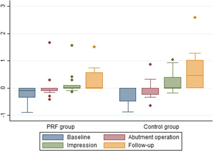
Figure 2. Box plot of the radiographic peri-implant marginal bone level at different time points in millimeter. Baseline: the time of implant placement; abutment: the time of abutment operation; impression: the time of impression taking; follow-up: the time of the final follow-up
Figure 2. Box plot of the radiographic peri-implant marginal bone level
author: Jens Hartlev,Sren Schou,Flemming Isidor, Sven Erik Nrholt | publisher: drg. Andreas Tjandra, Sp. Perio, FISID

Serial posts:
- A clinical and radiographic study of implants placed in autogenous bone grafts
- Background: A clinical and radiographic study of implants (1)
- Background: A clinical and radiographic study of implants (2)
- Material & methods: A clinical and radiographic study of implants (1)
- Material & methods: A clinical and radiographic study of implants (2)
- Material & methods: A clinical and radiographic study of implants (3)
- Material & methods: A clinical and radiographic study of implants (4)
- Material & methods: A clinical and radiographic study of implants (5)
- Material & methods: A clinical and radiographic study of implants (6)
- Results: A clinical and radiographic study of implants (1)
- Results: A clinical and radiographic study of implants (2)
- Results: A clinical and radiographic study of implants (3)
- Results: A clinical and radiographic study of implants (4)
- Discussion: A clinical and radiographic study of implants (1)
- Discussion: A clinical and radiographic study of implants (2)
- Discussion: A clinical and radiographic study of implants (3)
- Abbreviations & References: A clinical and radiographic study of implants
- Table 1 Demographics and survival rates of implants and implant crowns
- Table 2 Radiographic peri-implant marginal bone level in mm
- Table 3 Radiographic marginal bone level and clinical recession on neighbouring tooth surface
- Table 4 Patient-related outcome measures at baseline and at the final follow-up
- Figure 1. Intraoperative photos illustrating bone harvesting
- Figure 2. Box plot of the radiographic peri-implant marginal bone level
- Figure 3. Data from the VAS of patient-related outcome measures at the time of mounting of the implant-supported crown and at the final follow-up of the PRF and control group