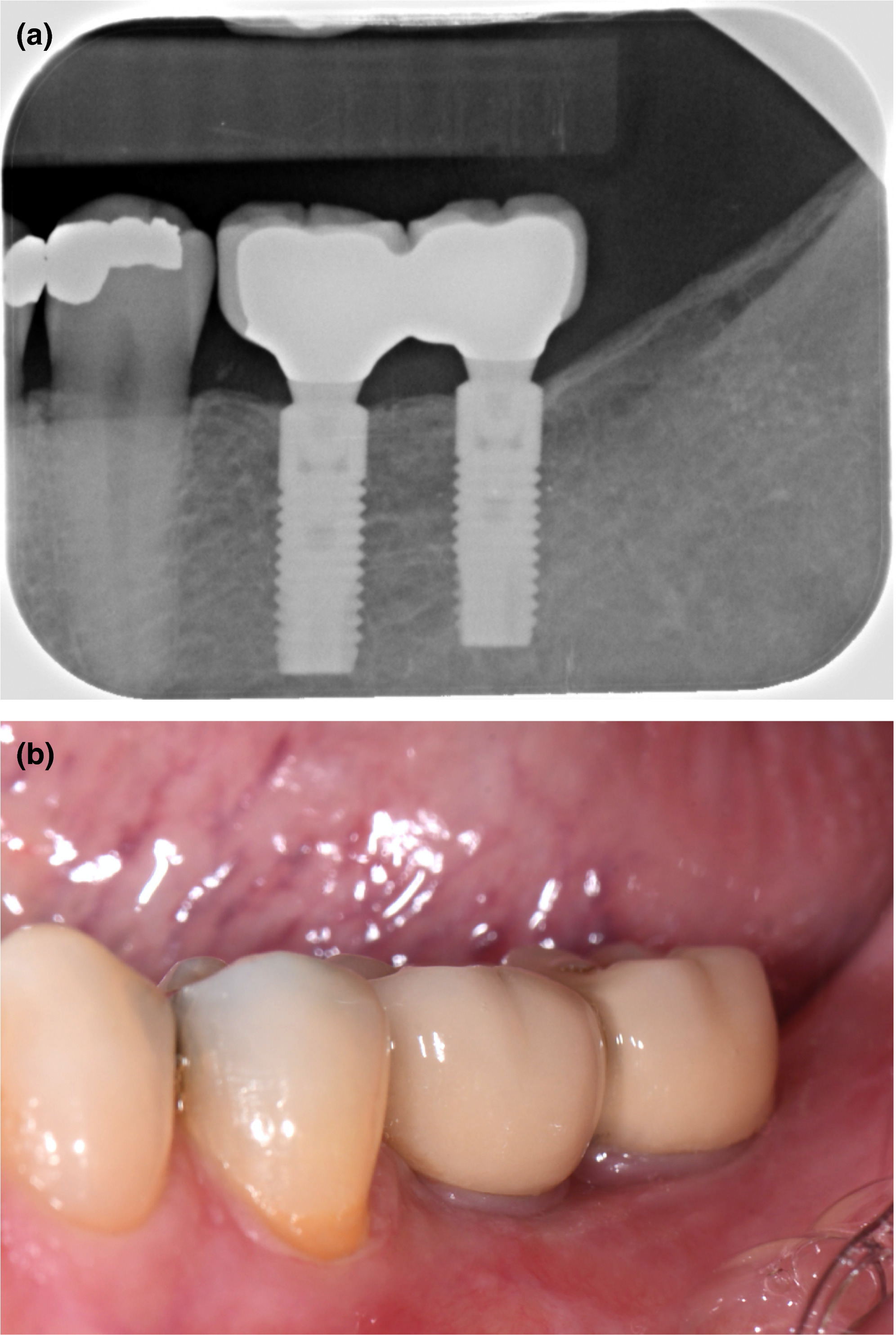
Figure 2. Five‐year follow‐up radiograph (a) and clinical photograph (b) of patient with two 11‐mm implants
Figure 2. Five‐year follow‐up of patient with two 11‐mm implants
author: Felix L Gulj,Henny J A Meijer,Ingemar Abrahamsson,Christopher A Barwacz,Stephen Chen,Paul J Palmer,Homayoun Zadeh,Clark M Stanfo | publisher: drg. Andreas Tjandra, Sp. Perio, FISID

Serial posts:
- Comparison of 6‐mm and 11‐mm dental implants in the posterior region
- Material & methods : Comparison of 6‐mm and 11‐mm dental implants (1)
- Figure 1: patient with two 6‐mm implants
- Figure 1a: Five‐year follow‐up radiograph of patient with two 6‐mm implants
- Figure 1b. Five‐year follow‐up clinical photograph of patient with two 6‐mm implants
- Figure 2. Five‐year follow‐up of patient with two 11‐mm implants
- Figure 2a. Five‐year follow‐up radiograph of patient with two 11‐mm implants
- Figure 2b. Five‐year follow‐up photograph of patient with two 11‐mm implants
- Material & methods : Comparison of 6‐mm and 11‐mm dental implants (2)
- Material & methods : Comparison of 6‐mm and 11‐mm dental implants (3)
- Material & methods : Comparison of 6‐mm and 11‐mm dental implants (4)
- Results : Comparison of 6‐mm and 11‐mm dental implants
- Table 1. Baseline characteristics
- Table 2. Mean value (in mm), standard deviation (SD), and frequency distribution
- Table 3. Clinical measures of implants
- Table 4. Number of technical complications at implant level and patient level
- DISCUSSION : Comparison of 6‐mm and 11‐mm dental implants (1)
- DISCUSSION : Comparison of 6‐mm and 11‐mm dental implants (2)
- DISCUSSION : Comparison of 6‐mm and 11‐mm dental implants (3)
- DISCUSSION : Comparison of 6‐mm and 11‐mm dental implants (4)
- CONCLUSIONS : Comparison of 6‐mm and 11‐mm dental implants