Figure 3: Pretreatment cephalometric radiograph and its tracing
Making space for missing middle incisor (13)
author: Andreas Tjandra | publisher: drg. Andreas Tjandra, Sp. Perio, FISID
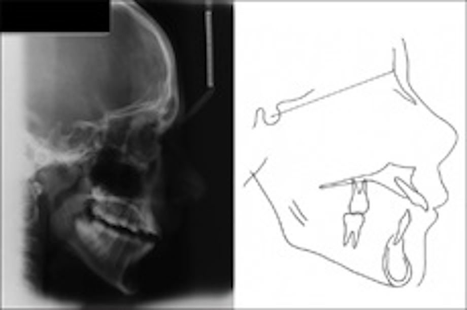
Figure 3:
Pretreatment cephalometric radiograph and its tracing
Serial posts:
-
A Comprehensive Analysis of Adult Tooth Removal Reasons (12)
-
Alasan Mencabut Gigi Dewasa (12)
-
IOPA Immediately after Crown cementation
-
Implants supported prosthesis in occlusion
-
Making space for missing middle incisor (13)
-
Do orthopedic treatments for growing retrognathic hyperdivergent patients lead to stable outcomes? (12)
Pretreatment cephalometric radiograph and its tracing
- A Comprehensive Analysis of Adult Tooth Removal Reasons (12)
- Alasan Mencabut Gigi Dewasa (12)
- IOPA Immediately after Crown cementation
- Implants supported prosthesis in occlusion
- Making space for missing middle incisor (13)
- Do orthopedic treatments for growing retrognathic hyperdivergent patients lead to stable outcomes? (12)