Case study, Peri-implantitis, Histomorphometry
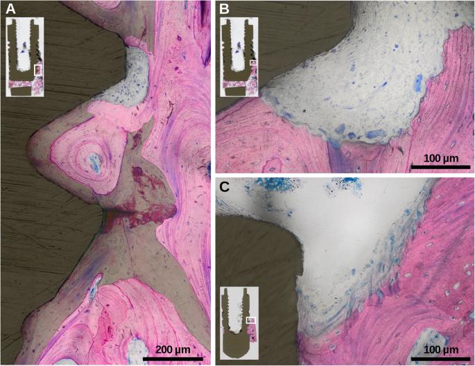
Fig. 3. Histological magnifications of specimens exposing a significant areas of lamellar bone in-between and parallel to the surface of older trabeculae (accentuated by green overlay), b osteoclastic activity, and c active bone formation at intrabony marginal regions : Histological characteristics of advanced peri-implant
author: Maria Elisa Galrraga-Vinueza, Stefan Tangl, Marco Bianchini, Ricardo Magini, Karina Obreja, Reinhard Gruber, Frank Schwarz | publisher: drg. Andreas Tjandra, Sp. Perio, FISID

Fig. 3. Histological magnifications of specimens exposing a significant areas of lamellar bone in-between and parallel to the surface of older trabeculae (accentuated by green overlay), b osteoclastic activity, and c active bone formation at intrabony marginal regions
Serial posts:
- Abstract : Histological characteristics of advanced peri-implantitis bone defects in humans
- Background : Histological characteristics of advanced peri-implantitis bone defects in humans
- Materials and methods : Histological characteristics of advanced peri-implantitis bone defects in humans [1]
- Materials and methods : Histological characteristics of advanced peri-implantitis bone defects in humans [2]
- Materials and methods : Histological characteristics of advanced peri-implantitis bone defects in humans [3]
- Results : Histological characteristics of advanced peri-implantitis bone defects in humans
- Discussion : Histological characteristics of advanced peri-implantitis bone defects in humans [1]
- Discussion : Histological characteristics of advanced peri-implantitis bone defects in humans [2]
- Conclusions : Histological characteristics of advanced peri-implantitis bone defects in humans
- Availability of data and materials : Histological characteristics of advanced peri-implantitis bone defects in humans
- Abbreviations : Histological characteristics of advanced peri-implantitis bone defects in humans
- References : Histological characteristics of advanced peri-implantitis bone defects in humans [1]
- References : Histological characteristics of advanced peri-implantitis bone defects in humans [2]
- References : Histological characteristics of advanced peri-implantitis bone defects in humans [3]
- References : Histological characteristics of advanced peri-implantitis bone defects in humans [4]
- Acknowledgements : Histological characteristics of advanced peri-implantitis bone defects in humans
- Funding : Histological characteristics of advanced peri-implantitis bone defects in humans
- Author information : Histological characteristics of advanced peri-implantitis bone defects in humans [1]
- Author information : Histological characteristics of advanced peri-implantitis bone defects in humans [2]
- Ethics declarations : Histological characteristics of advanced peri-implantitis bone defects in humans
- Additional information : Histological characteristics of advanced peri-implantitis bone defects in humans
- Rights and permissions : Histological characteristics of advanced peri-implantitis bone defects in humans
- About this article : Histological characteristics of advanced peri-implantitis bone defects in humans
- Table 1 Patient and implant site characteristics : Histological characteristics of advanced peri-implantitis bone defects in humans
- Table 2 Results from histomorphometric measurements exhibiting DL, RB, bone density (%), residual bone-to-implant contact (%) values : Histological characteristics of advanced peri-implantitis bone
- Table 3 Mean osteocyte (OD) and empty lacunae density (ELD) at peri-implant residual bone : Histological characteristics of advanced peri-implantitis bone defects in humans
- Fig. 1. Histological section illustrating the landmarks to determine the conducted histomorphometrical length measurements: DL, RB, and BIC at buccal RB. The red square frames the intra-thread area (ROI) for OD and ELD analysis : Histological characteristics of advanced peri-implant
- Fig. 2. Histological view (buccal aspect) displaying mean BIC (%) and mean bone density (%) at corresponding regions (500 μm zones) from BD to A. Blue lines along the implant perimeter display BIC presence and yellow lines display BIC absence : Histological characteristics of advanced peri-implant
- Fig. 3. Histological magnifications of specimens exposing a significant areas of lamellar bone in-between and parallel to the surface of older trabeculae (accentuated by green overlay), b osteoclastic activity, and c active bone formation at intrabony marginal regions : Histological characteristics of advanced peri-implant