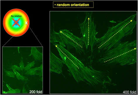
Figure 4. Randomly orientated osteoblasts without influence of rotation (phallacidin fluorescence staining). On the left side with 200× and on the right side with 400× magnification. The white X on the coloured circle marks the location upon the plate where the osteoblasts were located. The red X marks the centre of the plate
Figure 4. Randomly orientated osteoblasts without influence of rotation
author: P W Kmmerer,D G E Thiem,A Alshihri,G H Wittstock,R Bader,B Al-Nawas, M O Klein | publisher: drg. Andreas Tjandra, Sp. Perio, FISID

Serial posts:
- Cellular fluid shear stress on implant surfaces
- Methods : Cellular fluid shear stress on implant surfaces (1)
- Methods : Cellular fluid shear stress on implant surfaces (2)
- Methods : Cellular fluid shear stress on implant surfaces (3)
- Results : Cellular fluid shear stress on implant surfaces (1)
- Results : Cellular fluid shear stress on implant surfaces (2)
- Discussion : Cellular fluid shear stress on implant surfaces (1)
- Discussion : Cellular fluid shear stress on implant surfaces (2)
- Discussion : Cellular fluid shear stress on implant surfaces (3)
- Discussion : Cellular fluid shear stress on implant surfaces (4)
- References : Cellular fluid shear stress on implant surfaces
- Figure 1. Three-dimensional illustration and photography
- Figure 2. Side view of a computerized simulation
- Figure 3. Diagram for visualisation of the calculation of shear stress rates
- Figure 4. Randomly orientated osteoblasts without influence of rotation
- Figure 5. Osteoblasts with an orientation tendency after 24 h