Introduction: Combination Syndrome
Introduction
Combination syndrome (CS) is defined as “a condition caused by the presence of the lower anterior teeth and the absence of the posteriors and resulting in significant maxillary anterior alveolar resorption.”1 This condition often develops in cases of a complete maxillary denture opposing a bilateral distal extension mandibular partial denture2 (Figures 1 - 3). The resulting chronic occlusal trauma from the mandibular anterior teeth to the premaxillary hard and soft tissue structures often leads to a slow resorption of the anterior maxillary alveolar ridge that is replaced with the fibrous tissue. Ellsworth Kelly followed up 6 patients for 3 years with a complete maxillary denture opposed by mandibular anterior teeth and a distal extension removable partial denture (RPD).3 Kelly gave the name “combination syndrome” to this condition. He described 3 key features of CS: reduction of anterior maxillary bone, enlargement of maxillary tuberosities, and bone resorption under the mandibular RPD bases. A lack of posterior occlusion and the presence of excessive anterior occlusal function by super-erupted anterior mandibular teeth led to the use of another name for this condition, “an anterior hyperfunction syndrome.”2 Other features that may be present at the same time include loss of vertical dimension of occlusion, occlusal plane discrepancy, anterior spacial reposition of the mandible, poor adaptation of the prosthesis, and others.4,5 Prevention of loss of posterior occlusion and avoidance of anterior hyperfunction are considered the main treatment approaches of this complex condition. An implant-based reconstruction of the mandibular arch with implant stabilization of the maxillary arch6 is a modern bi-maxillary surgical-prosthetic therapy for many CS cases.
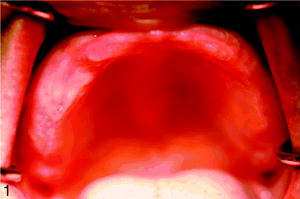
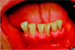
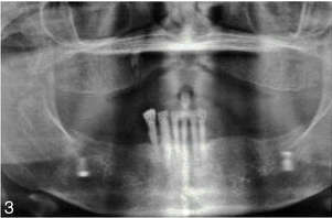
Severe maxillary resorption is a dominant feature of combination syndrome patients. These CS patients as well as many other clinical cases of extensive three-dimensional maxillary bone loss have guided dental clinicians and surgeons towards the development of many innovative surgical and prosthetic techniques of correction of maxillary bone atrophy combined with immediate or delayed placement of dental implants. These reconstructive approaches for the compromised maxillary bone include vertical alveolar distraction osteogenesis,7,8 horizontal distraction in combination with bilateral sinus lift/bone grafting procedure,9,10 maxillary ridge-splitting techniques followed by immediate dental implants,11 autogenous iliac crest12 and calvarial bone grafting,13 reconstruction of the resorbed edentulous maxilla with autogenous rib grafts,14 tibial grafting for maxillary bone loss,15 treatment of severe maxillary atrophy with vascularized free fibula flap in combination with dental implants,16 interpositional bone grafting with LeFort I osteotomy,17 orthognathic surgery with or without onlay bone grafting,18,19 use of the osseoinductive effect of bone morphogenic protein within endosseous dental implants placed in the maxilla,20 zygomatic implants with or without sinus lift/bone graft,21,22 pterygomaxillary implants combined with zygomatic and conventional implants,23 the Marius implant bridge for the surgical-prosthetic rehabilitation of the resorbed completely edentulous maxilla with 6 implants,24 “all-on-4” maxillary edentulous rehabilitation with 4 strategically placed and immediately loaded implants,25 combination of short implants and osteotome technique for the posterior maxilla,26 use of transitional implants and bone grafting before placement of definitive implants,27 optimal use of the anatomic features of the maxillary arch with tilted implants,28 use of transmandibular implant29 and Tatum custom ramus frame implant in CS patients,30 and others.
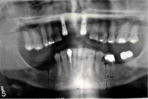
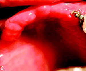
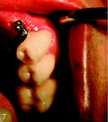
In typical CS cases, a maxillary ridge is completely edentulous. However, patients with a partial maxillary edentulism can also have similar signs and symptoms. Cases of maxillary RPD with an anterior edentulous space and preserved posterior teeth opposed by mandibular anterior teeth and a distal extension RPD or natural mandibular dentition demonstrate analogous deteriorating effects of CS. It appears that when the posterior maxillary teeth present on one or both sides and opposed by the posterior mandibular teeth, the hypertrophic changes (overgrowth) of the posterior maxilla and maxillary tuberosities are not prevalent or do not develop at all. Due to preservation of the posterior occlusal relationship, the anatomy of posterior quadrants of both jaws is not altered significantly in these cases (Figures 4 and 5). In Figures 4 and 5, the hypertrophic changes on the right posterior maxilla and super-eruption of unopposed posterior molars are more pronounced than the changes on the left side, where the posterior occlusal relationships between periodontally healthy upper and lower teeth remain.
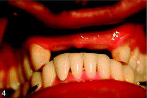
Both implant-retained and implant-supported prostheses have become an increasingly popular and successful prosthetic rehabilitation for partially and fully edentulous maxilla.31,32 This case report demonstrates an implant treatment of the partially edentulous maxilla with bilaterally preserved posterior teeth opposed by a fixed crown and bridge mandibular dentition in the patient with typical features of CS. Dental implants together with natural teeth were used for retention of an implant-assisted maxillary overdenture. A new classification of combination syndrome is proposed in this article following the case report.
Serial posts:
- Combination Syndrome: Classification and Case Report
- Introduction: Combination Syndrome
- Case report: combination syndrome
- Diagnosis: Combination Syndrome
- Treatment Plan : Combination Syndrome
- Operative phase : Combination Syndrome
- Restorative stage : Combination Syndrome
- Classification : Combination Syndrome
- Discussion : Combination Syndrome
- Conclusion : Combination Syndrome
- References : Combination Syndrome
- Table Classification of combination syndrome (CS)
- Extended table of classification of Combination Syndrome (CS)